
Presentation
This male patient has respiratory failure and is awaiting a lung transplant. He presented with an infective exacerbation of his disease.
Patient Data
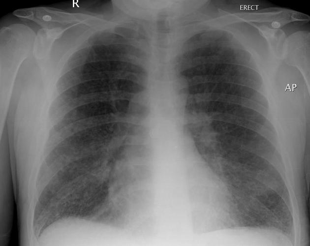
The plain frontal chest x-ray shows mild hyperinflation and a coarse pattern in the lung fields consisting of reticular interstitial markings and peripheral ring shadows suggesting cysts.
Case Discussion
High-resolution CT confirmed the presence of multiple small peripheral cystic lesions, some of which were branching and bizarre. There was honeycombing in the lower zones. There were a few small peribronchovascular nodules and there was interlobular septal thickening. The differential diagnosis of multiple pulmonary cystic lesions includes Langerhans cell histiocytosis, lymphangioleiomyomatosis, sarcoidosis and lymphocytic interstitial pneumonitis. Peripheral bronchiectasis should be considered as it may appear cystic. Given the other changes seen on HRCT and the patient’s sex, LCH was the favored imaging diagnosis.
Image contributed by: Dr Laughlin Dawes


 Unable to process the form. Check for errors and try again.