
Presentation
Known case of chronic kidney disease with uncontrolled diabetes presenting with cough, fever and breathlessness.
Patient Data
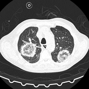

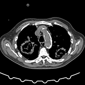

Multiple bilateral rounded areas of reticulation and ground glass opacity surrounded by denser outer rings (bird’s nest appearance).
Bronchial wall thickening and plugging in the posterior lower lobes. Collapse and consolidation most marked in the right lower lobe.
Small bilateral pleural effusions.
Case Discussion
The features suggest invasive fungal disease such as Mucormycosis which is rapidly progressive and resistant to conventional antifungals. Identification of the organism often requires lung biopsy. When disease is localized, excision of the affected tissue may be life-saving.
Angioinvasion leads to pulmonary infarction and centrifugal invasion of contiguous tissue creating the bird’s nest appearance.
Mucormycosis occurs in immunocompromised patients including diabetics.


 Unable to process the form. Check for errors and try again.