
Presentation
Shortness of breath. Echocardiography showed signs of pulmonary arterial hypertension.
Patient Data
Age: 45 years
Gender: Female
From the case:
Pulmonary thromboembolism on MRI


Download
Info
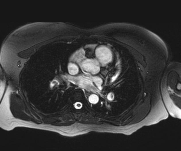
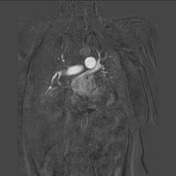
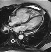
Filling defects, likely thromboemboli, are demonstrated within both pulmonary arteries.
The right ventricle is enlarged with bowing of the interventricular septum towards the left ventricle reflective of right ventricular strain.
Minimal pericardial effusion is seen.
Incidentally, there are hepatic cysts.
Case Discussion
This is a case of pulmonary thromboembolism with right ventricular strain demonstrated on magnetic resonance imaging (MRI).


 Unable to process the form. Check for errors and try again.