
Presentation
Primary amenorrhea.
Patient Data
Age: 25 years
Gender: Female
From the case:
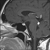
Quadrigeminal cistern lipoma

Download
Info

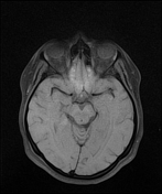
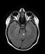
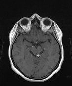
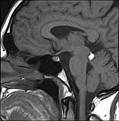
The MR sequences demonstrate a small well-defined lesion in the left para-median region of the quadrigeminal cistern. It shows high intensity on both T1 and T2 with loss of signal on axial T1 fat saturation.
The pituitary gland is normal in size with no focal lesion seen.
Case Discussion
MRI features characteristic of a quadrigeminal cistern lipoma.
The differential diagnosis should include:
- subarachnoid hemorrhage: gradient echo and fat-saturated sequences are the best sequences to differentiate it from a lipoma
- intracranial fat-containing lesions: see related article intracranial lipoma


 Unable to process the form. Check for errors and try again.