
Presentation
Incidental finding.
Patient Data


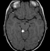
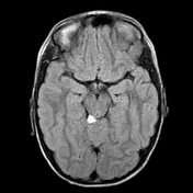

MRI of the brain demonstrates a rounded region of intense intrinsic high T1 signal located on the right, in the quadrigeminal plate cistern, abutting the inferior colliculus. On FLAIR and T2 weighted sequences it is also of high signal, with subtle chemical shift artefact seen on T2. On fat-saturated sequences, it fully attenuates.

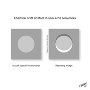
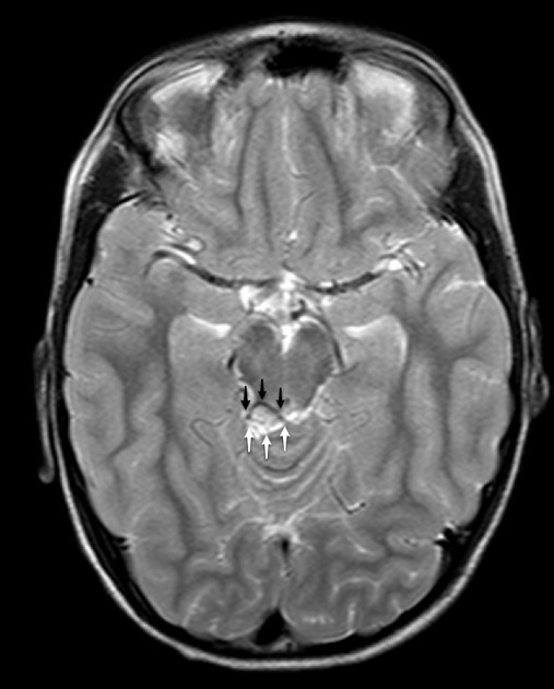
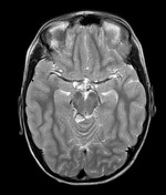
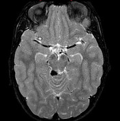
T2 weighted image demonstrates chemical shift artifact around the lipoma in the frequency encoding direction (anteroposterior). Note how there is signal loss anteriorly (black arrows) and hyperintensity posteriorly (white arrows). This is the result of inaccurate spatial encoding, resulting in the lipoma being incorrectly placed posterior to its actual location.
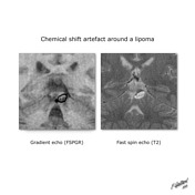
It should be noted that this appearance is not seen on gradient-echo sequences, where signal drops out will be in all directions.
Case Discussion
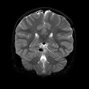
This case demonstrates typical appearances of a quadrigeminal cistern lipoma, with prominent chemical shift artifact visible on T2 weighted fast spin-echo images.


 Unable to process the form. Check for errors and try again.