Presentation
Headache.
Patient Data
Age: 20 years
Gender: Male
From the case:
Quadrigeminal cistern lipoma
![](https://prod-images-static.radiopaedia.org/images/61444455/98aaf38cfb7069138114a9c55fecd815e5f32f7e5a5f99a697ee7ac3ac342a0d_thumb.jpeg)
![This study is a stack](/packs/stack-YQKLCKBI.gif)
![](https://prod-images-static.radiopaedia.org/images/61444473/a530d1f1cd91c413b82b56162ac7ab21622afafaa468a099faef2171ee4cb1a1_thumb.jpeg)
![This study is a stack](/packs/stack-YQKLCKBI.gif)
![](https://prod-images-static.radiopaedia.org/images/61444491/e725a379cf030681b84ab73a1e37dea676044598f622feaf0b07a88446601479_thumb.jpeg)
![This study is a stack](/packs/stack-YQKLCKBI.gif)
![](https://prod-images-static.radiopaedia.org/images/61444611/bac0bba3e274641c64c7c3ec75cac976f4ce15c5875568106c150f2f16ef3ad7_thumb.jpeg)
![This study is a stack](/packs/stack-YQKLCKBI.gif)
![](https://prod-images-static.radiopaedia.org/images/61444519/1dac76fc0ab1076144938c1980dfef8cacefd3fc4071fe8a24879ea31fc43846_thumb.jpeg)
![This study is a stack](/packs/stack-YQKLCKBI.gif)
![](https://prod-images-static.radiopaedia.org/images/61444561/8bc540c1d3006fe2c4dca8fe45e29d1a21c08c7332324e71ab2802449ec50680_thumb.jpeg)
![This study is a stack](/packs/stack-YQKLCKBI.gif)
![](https://prod-images-static.radiopaedia.org/images/61444536/808c257d6ef27b3fd2f2f217e70476517d0fc3bc31165c4f67e5f4aa97894fa8_thumb.jpeg)
![This study is a stack](/packs/stack-YQKLCKBI.gif)
Download
Info
![](https://prod-images-static.radiopaedia.org/images/61444455/98aaf38cfb7069138114a9c55fecd815e5f32f7e5a5f99a697ee7ac3ac342a0d_big_gallery.jpeg)
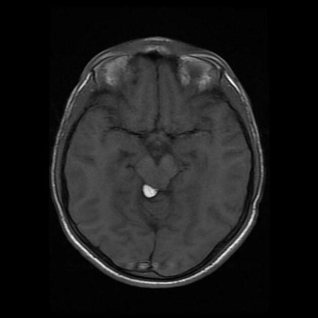
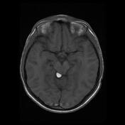
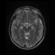
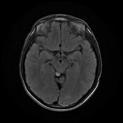
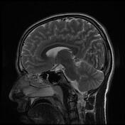
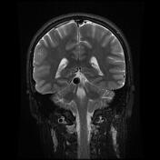
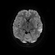
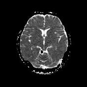
A well-defined fat equivalent signal lesion is seen on the right side of the quadrigeminal cistern just posterior to the tectum/midbrain with a minimal mass effect that shows a signal drop on fat suppression sequences, in keeping with quadrigeminal lipoma.
Mild mucosal thickening is present in paranasal sinuses.
Case Discussion
MRI features are characteristic of a quadrigeminal cistern lipoma which is the second commonest site for intracranial lipomas after pericallosal lipoma.