
Note: This case has been tagged as "legacy" as it no longer meets image preparation and/or other case publication guidelines.
From the case:
Quadrigeminal plate lipoma


Download
Info

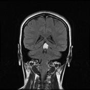
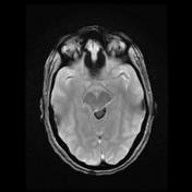
A mass in the quadrigeminal plate cistern, just below the pineal gland has high T1 and T2 signal and attenuates on fat saturated T1 C+ sequence. Additionally, it demonstrates prominent chemical shift artifact on the sagittal T2 and gradient-echo sequences. Features consistent with a lipoma.
Case Discussion
Appearances are typical of an intracranial lipoma. If fat saturated sequences are not available, then presence of chemical shift artifact is helpful in confirming the mass is fatty.


 Unable to process the form. Check for errors and try again.