
Presentation
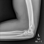
Left elbow pain, since 5 weeks after falling down during playing football.
Patient Data

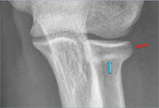

There is evidence of faintly seen discontinuity and cortical step-off (red arrow) involving the lateral aspect of the radial head, and impaction line or sclerotic band (blue arrow) involving the neck of the radius representing radial head fracture.
According to Mason classification of radial head fractures representing type I.
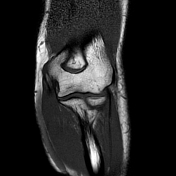

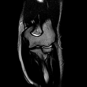


The provided MRI sequences revealed an abnormal hypointense irregular line on T1-weighted images and high signal intensity on T2-weighted images and STIR sequences, involving the neck of the radius, Features are going with radial head fracture and minimal impaction, associated with diffuse bone marrow edema.
Case Discussion
The radial head fracture should be assessed carefully to avoid missing subtle fractures.
In general, for looking after fractures, it advised following these instructions on radiographs:
sometimes one view is not enough to detect a fracture, so ask for another view
magnification of the images is recommended and follows the cortex as well
direct signs of fracture like break of discontinuity of the cortex, cortical step-off, bony fragment and impaction line or sclerotic band
indirect signs of fracture like a displaced fat pad, joint effusion and soft tissue swelling should also be assessed
Co-author: Ahmed Mostafa Hassan Wahdan MD, Radiologist


 Unable to process the form. Check for errors and try again.