
Patient Data
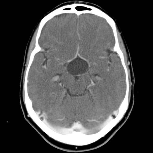
Contrast-enhanced CT images showing a cystic lesion within the sella and suprasellar area. No contrast-enhancement or wall thickness.

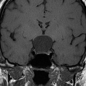
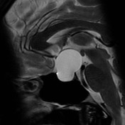
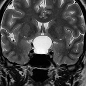
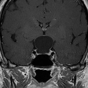
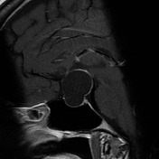
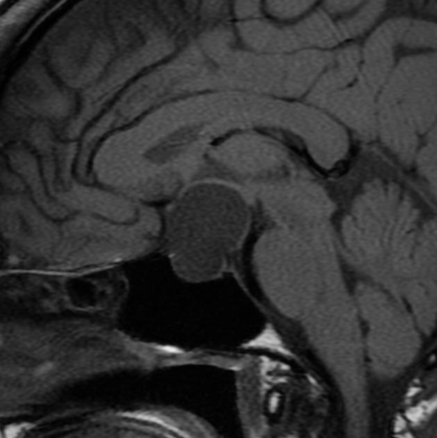
Selected MRI images demonstrate a large sellar and predominantly suprasellar cyst characterized by thin walls and homogeneous content similar to the CSF signal.
Case Discussion
The patient went on to have a resection.
Histology:
Gross Description: The specimen is received in a single container labeled with the patient's demographics and SUPRASELLAR CYST. It consists of multiple fragments of thin translucent membranous tissue.
Microscopic Description: Sections show fragments of a cyst wall lined by a continuous layer of benign cuboidal epithelium. In several fragments, normal anterior pituitary is intimately associated with the cyst wall. In other fragments, the cyst wall is supported by hypocellular fibrous connective tissue. The histologic appearance is consistent with Rathke's cleft cyst.
FINAL DIAGNOSIS: cyst consistent with Rathke's cleft cyst.


 Unable to process the form. Check for errors and try again.