
Presentation
Chronic constipation.
Patient Data
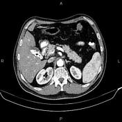

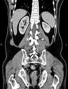
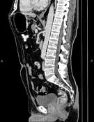

Multiple stones are seen in gallbladder less than 18mm.
A few non-enhanced simple cortical cysts are seen at both kidneys, with maximum diameters of 25mm. In addition, a few small stones less than 4mm are noted in kidneys.
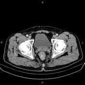
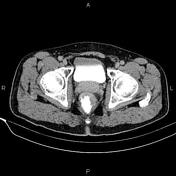
Asymmetrical increased wall thickness suggestive of tumoral infiltration is present in the distal rectum. There is mild perirectal fat stranding and a few small lymph nodes are also evident.
The prostate gland is enlarged.
Degenerative changes as osteophytosis are seen at the lumbar spine.
Case Discussion
Pathology proven rectal adenocarcinoma with small regional lymph nodes. Colorectal cancers can be found anywhere from the cecum to the rectum. Rectosigmoid involvement includes about 55% of cases as the most common site of colorectal cancer. CT is the modality most used for staging colorectal carcinoma, however, MRI is the preferred modality for the staging of rectal cancer.


 Unable to process the form. Check for errors and try again.