
Presentation
Right upper arm pain and swelling. History of previous curettage of a bony mass lesion in that location.
Patient Data
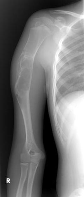
A well-defined right proximal humeral diaphyseal expansile, multilocular lesion with a geographic, sclerotic margin. No matrix calcification and no visible periosteal reaction.


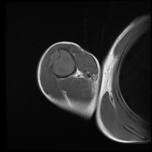
An expansile, mildly lobulated, septated lesion is seen within the humeral diaphysis. The lesion shows low-to-intermediate T1 and high T2/STIR signal intensity with a T1/T2 hypointense sclerotic rim. The lesion measures 11 x 3 x 3 cm in its main CC, TS and AP axes, respectively. There is endosteal scalloping with a sclerotic margin and a narrow zone of transition. Focal cortical destruction is noted with a small extraosseous soft tissue component at the anterolateral aspect of the lesion. No fluid/fluid levels are seen. The lesion shows heterogeneous post-contrast enhancement, with enhancing septa.
Case Discussion
Presumed recurrent chondromyxoid fibroma of the proximal humerus.
The differential diagnosis is:
- Aneurysmal bone cyst (ABC): fluid/fluid level, periosteal reaction, no matrix mineralization. Chondromyxoid fibroma with secondary ABC is not uncommon.
- Fibrous dysplasia: central location, no internal septation or periosteal reaction. Ground glass attenuation is typical.
- Adamantinoma: older patient, vast majority in the upper tibia, cortical destruction with aggressive periosteal reaction.
- Fibrous cortical defect (FCD) and non-ossifying fibroma (NOF): Possible mild cortical expansion, no destruction or periosteal reaction, not painful.


 Unable to process the form. Check for errors and try again.