
Presentation
Left flank pain. Fever. Ultrasound suggested left kidney mass. Abscess?
Patient Data
Age: 50 years
Gender: Female
From the case:
Renal abscess with CT guided drainage
Download
Info
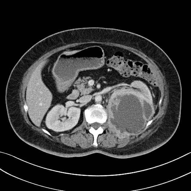
Large left-sided renal abscess displacing the remaining or the kidney anteriorly. No renal stones.
Gallstones. No free fluid.


Download
Info

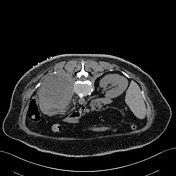
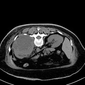
Percutaenous drainage requested. Unable to undertake with US
Prone position.
The abscess was entered with an access needle.
Seldinger technique.
12F drain inserted.
Frank pus drained freely.
Download
Info
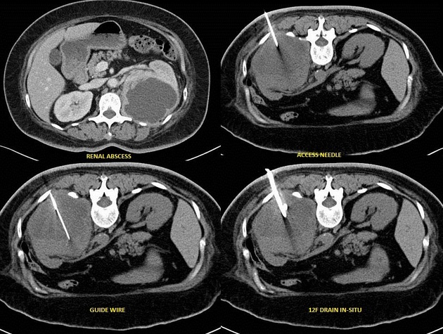
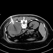
The key steps in the Seldinger process illustrated.
Case Discussion
CT may be required for drainage procedures.
Reasons include:
- size of patient
- depth of the collection
- unable to get a safe window for drainage
- inadequate visualization (i.e collection of mixed echogenicity)
Unlike ultrasound where a direct puncture is an option for ultrasound-guided drainage, CT requires the Seldinger technique for drain insertion.


 Unable to process the form. Check for errors and try again.