
Presentation
Left flank pain
Patient Data
Age: 65 years
Gender: Female
From the case:
Renal angiomyolipoma


Show annotations
Download
Info
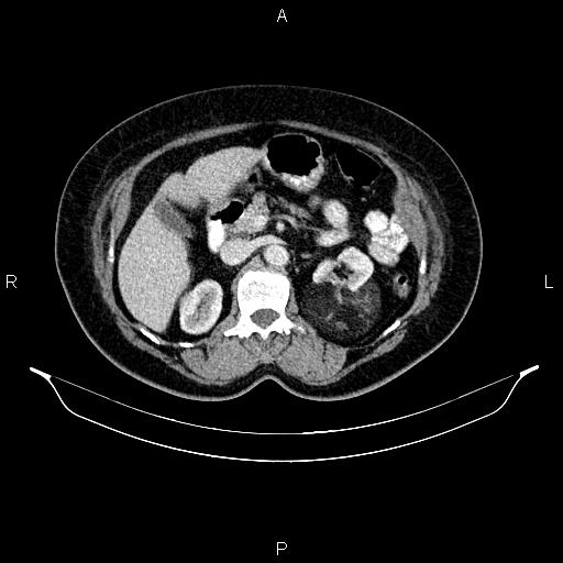
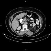
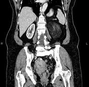
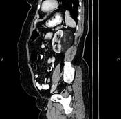
A 105 × 70 × 75 mm exophytic well-defined fatty mass extrudes from the posterior aspect of the left kidney and maintains broad contact with the capsule (overflowing beer sign). Several vessels course through the fat.
Degenerative changes such as osteophytosis are seen in the lumbar spine.
Case Discussion
Features are typical for left renal angiomyolipoma, which is a type of benign renal neoplasm encountered both sporadically and as part of a phakomatosis, most commonly tuberous sclerosis.
On CT scan images most lesions involve the cortex and demonstrate macroscopic fat (less than -20 HU).


 Unable to process the form. Check for errors and try again.