
Presentation
Acute right flank pain
Patient Data


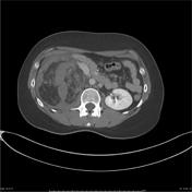
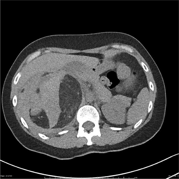
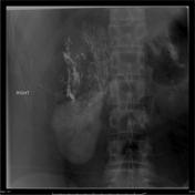
Heterogeneous fat containing rounded exophytic mass extending off the upper pole of the right kidney measuring 11.7 x 10.7 x 15.4 cm. The angiomyolipoma demonstrates internal density consistent with hematoma and hematoma also seen in the right pararenal space extending particularly posterior and inferior to the kidney, the largest portion measuring approximately 8.7 x 3.2 x 6.6 cm. The right kidney is displaced anteriorly and inferiorly as have the gallbladder and pancreatic head. The IVC is partially compressed. Small amount of free fluid seen around the liver. No renal calculus. No calculi seen in the path of the ureters. The liver, spleen, left adrenal gland and bowel are unremarkable. The right adrenal gland is not well discerned.
Contrast blush is seen on the delayed phase images in multiple places on the
lateral margin of the angiomyolipoma (seen on thin sections, not shown). Assessment for active extravasation in the center of the lesion is
difficult given the small caliber arteries.
The IVC is displaced anteriorly and compressed by mass-effect from essentially complete effacement at the T12/L1 level extending to the region of hepatic vein insertion.
Non-occlusive filling defects are seen within the left renal vein, superior azygos vein, left common iliac vein and inferior IVC.
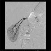

Initial angiographic runs showed a significantly ptosed right kidney, with supply to the large upper pole angiomyolipoma via tortuous artery arising from the superior division of the right renal artery.
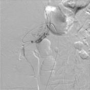
Selective catheterization of the branch supplying the angiomyolipoma was achieved via a microcatheter, and contrast runs through this vessel showed tumor filling, but no supply to renal parenchyma. Embolization was performed using a 50:50 dilution of absolute alcohol in lipiodol. A total of 12 mL was injected under careful angiographic observation to ensure no reflux to vessels supplying renal cortex.
Conclusion: Angiographically successful alcohol embolization of the right renal angiomyolipoma.


 Unable to process the form. Check for errors and try again.