
Presentation
Persistent left knee pain not responding to simple analgesia. No past medical history.
Patient Data

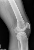
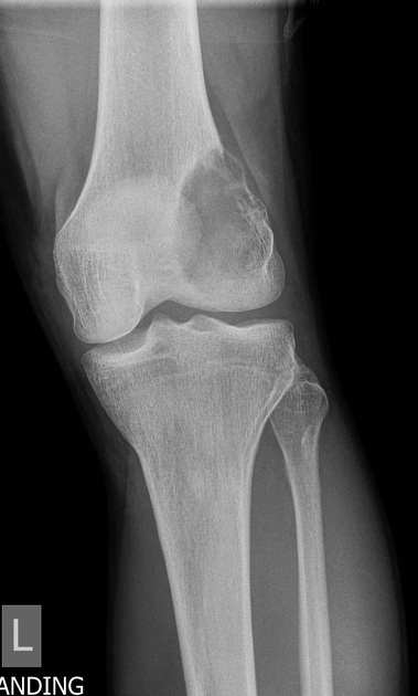
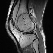
Lucent expansile lesion in the lateral femoral metaphysis with cortical thinning and breech.
No periosteal reaction.

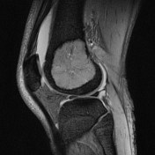
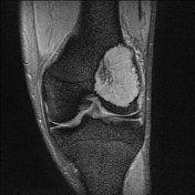
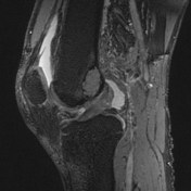
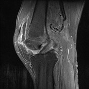

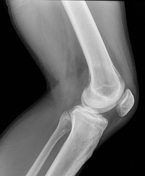
Lucent expansile lesion in the lateral femoral epiphysis and metaphysis with cortical breach.
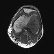
It exhibits intermediate T1 and high T2 signal with mild contrast enhancement.
Small joint effusion.
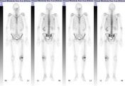


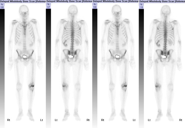
High tracer uptake in the distal femur corresponding with the other modalities.
CT of the chest, abdomen and pelvis identified a renal mass.
Subsequent biopsy of the femoral lesion indicated a renal cell carcinoma metastasis.
Case Discussion
A common site of metastasis of renal cell carcinoma is the skeleton, giving rise to lytic expansile metastases.
On occasion, the first presentation (presenting symptoms) may be from a bony metastasis. In this case, the lesion was thought to initially represent a primary bone lesion, which is within the differential.
Solitary bone lesions may be broadly divided into aggressive and non-aggressive from their appearances on plain film.


 Unable to process the form. Check for errors and try again.