
Presentation
History of left eye surgery 35 years ago. No details are available. On background of poor vision for 35 yrs, the patient presented with acute loss of vision for 1 month. USG to check for retinal detachment and ? IOL.
Patient Data

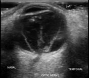
Globe - normal size and contour. The axial length is measured at 23 mm
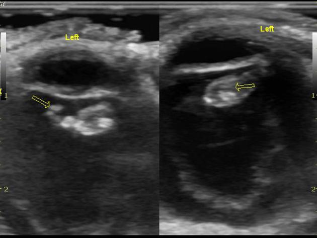
Lens - No intraocular lens is seen. Instead, altered contour with echogenic material in location of lens.
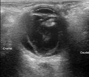
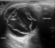
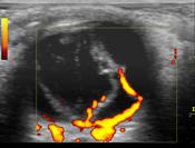
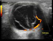
A 'V' shaped thick membrane is noted in all quadrants. No after movements are noted in this membrane, indicating retinal detachment.
Vitreous membranes are noted with 'v' shaped thick membrane. They show 'after movements'.
Another thick membrane is noted outer to retinal detachment on temporal side, indicating choroidal detachment. No after movements are noted in this membrane.


 Unable to process the form. Check for errors and try again.