
Presentation
Neutropenia during consolidation chemotherapy for acute myeloid leukemia. Presents with right preseptal painful swelling extending to the nose and reduction in right-sided visual acuity and hemianopia.
Patient Data


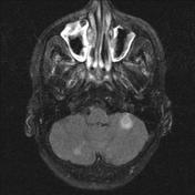


There is necrosed mucosa in the right nasal cavity and the right ethmoid sinuses, which is typically seen in acute invasive fungal sinusitis; the most common organisms in the given clinical context are mucor or Aspergillus.
Post-contrast sequences show non-enhancing angioinvasive fungal infection in the mucosa that gives the "black turbinate sign" appearance.
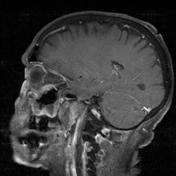
Within the brain, areas of diffusion restriction are in keeping with acute infarcts in the cerebellar hemispheres, bilateral medial occipital lobes, lateral aspect of the right thalamus and the white matter surrounding the left occipital horn.
There is an associated leptomeningeal enhancement indicative of meningitis, primarily in the medial occipital sulci.
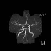
The left posterior cerebral artery shows distal vessel irregularity, with attenuation of the P3 branches before distal occlusion in the area of diffusion restriction.
Case Discussion
This case demonstrates the MRI appearances of acute invasive fungal sinusitis, together with one of its dreaded complications: septic infarction.


 Unable to process the form. Check for errors and try again.