
Presentation
Epigastric pain.
Patient Data
Age: 45 years
Gender: Female
From the case:
Riedel lobe


Download
Info
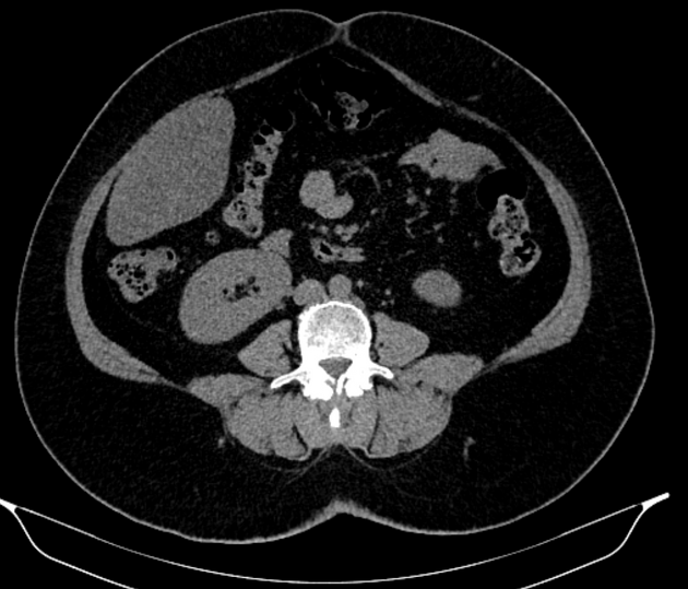
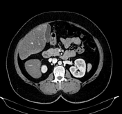
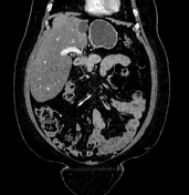
Tongue like inferior projection of the right lobe (segment 6) of the liver beyond the level of the most inferior costal cartilage.
An exophytic, non-enhancing cystic lesion with internal septae and tiny wall calcification arising from segment 2 of the liver. Imaging differentials include biliary cystadenocarcinoma, biliary cystadenoma, or hydatid cyst. Biopsy advised.
Diffuse hepatic steatosis: diffuse decrease in liver attenuation with absolute attenuation of 37 HU on contrast-enhanced scan. In non-enhanced images, the attenuation of the liver (11 HU) is much lower in comparison to the spleen (37HU) with a difference of >10 HU.
Case Discussion
Riedel lobe is an anatomical variant of the liver.


 Unable to process the form. Check for errors and try again.