
Presentation
Recent endotracheal tube placement.
Patient Data
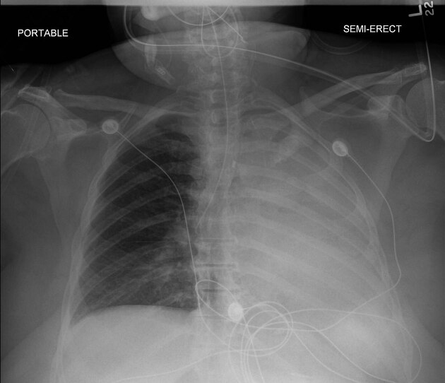
Initial chest x-ray shows an endotracheal tube in the right main bronchus/bronchus intermedius with complete opacification left hemithorax and shift of the mediastinum to the left indicating complete atelectasis left lung. NG tube coiled in the hypopharynx/proximal oesophagus.
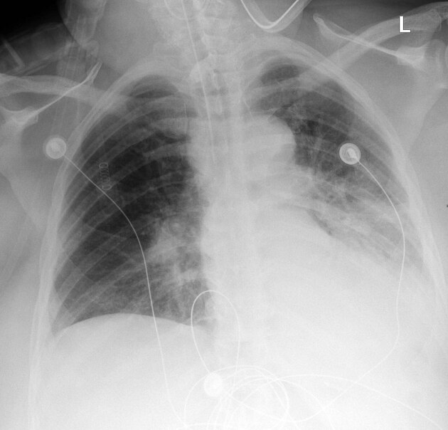
Subsequent chest x-ray after retraction of the endotracheal tube shows tube tip appropriately positioned above the carina and partial reexpansion left lung. Both studies show a nasogastric tube coiled in the pharynx/proximal oesophagus.
Case Discussion
The tip of an endotracheal tube should be positioned above the carina. If positioned too far distally, the tip most often ends up in the right bronchus because the right bronchus has a more direct origin from the trachea than the left bronchus. Tube placement into the right bronchus often results in atelectasis of the left lung which can be "cured" by retracting the tube proximal to the carina.


 Unable to process the form. Check for errors and try again.