
Presentation
The patient was admitted for preparation for herniated disc surgery, and a chest X-ray incidentally revealed the finding.
Patient Data
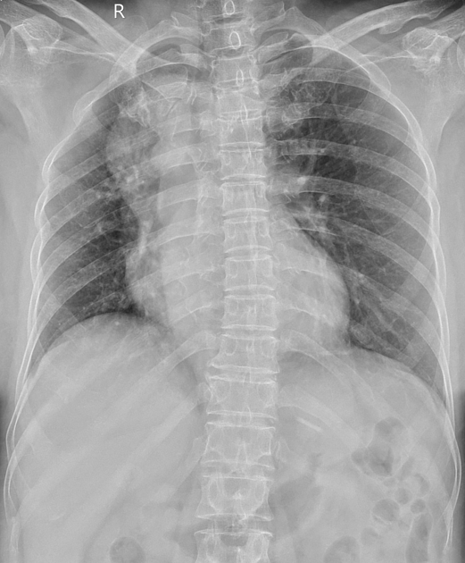
On radiograph:
the descending aorta and the aortic arch are located on the right, appearing elevated and projecting outward as a mass-like structure adjacent to the right side of the trachea
the trachea is deviated to the right


Right aortic arch with a Kommerell's diverticulum at the origin of an aberrant left subclavian artery with the diverticulum orifice diameter approximately 22 mm. The common carotid artery and right subclavian artery arise from the aortic arch. Calcification of the aortic arch, left subclavian artery, and coronary artery (LAD branch) is also observed. No evidence of dissection or thrombus in the vessel lumen.
Several areas of ground-glass opacities with associated bilateral interstitial thickening are also observed in the lung parenchyma.
Case Discussion
Right aortic arch with an aberrant left subclavian artery and a Kommerell's diverticulum.
This patient was incidentally detected, with no clinical symptoms and no evidence of compression, rupture, or dissection on imaging; thus, no further intervention has been undertaken.


 Unable to process the form. Check for errors and try again.