
Presentation
Sacral pressure ulcer.
Patient Data
Age: 55 years
Gender: Female
From the case:
Sacrococcygeal osteomyelitis and abscess


Download
Info
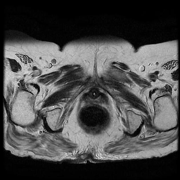
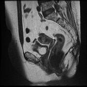
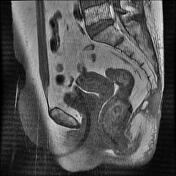
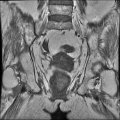
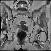
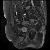
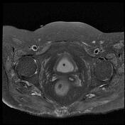
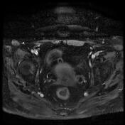
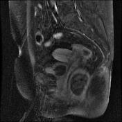
30 x 40 x 55 mm midline rim enhancing fluid signal collection associated with edema throughout the lower gluteal region superficial to the coccyx (first and second coccygeal segments) and distal sacrum (S1).
An enhancing sinus tract is present that terminates in the lower intergluteal cleft distal to the level of the last coccygeal segment.
Decreased T1 and hyperintense T2 fat suppression signal present throughout the remainder of the coccyx (with destruction) and in the S5 body.
Case Discussion
The MRI imaging findings represent osteomyelitis of sacrococcygeal and pericoccygeal soft tissue collection.


 Unable to process the form. Check for errors and try again.