
Presentation
Right visual loss. Had surgery for retinal detachment 10 years ago.
Patient Data
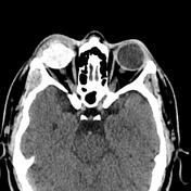

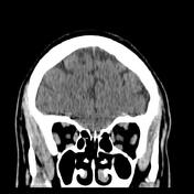
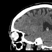
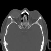
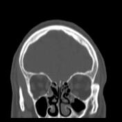
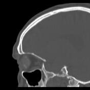
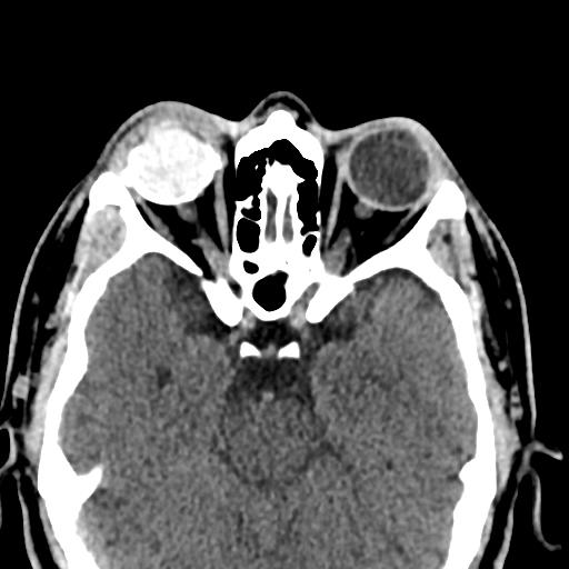
Small atrophied and retracted right ocular globe with dystrophic calcification in keeping with a phthisis bulbi. Spontaneously hyperdense vitreous humour (vitreous haemorrhage).
A hyperdense ring encircling the right ocular globe (previous history of scleral buckle surgery).
Case Discussion
CT features of a phthisis bulbi in a patient who had a scleral buckle surgery for retinal detachment with vitreous haemorrhage 10 years prior.
Phthisis bulbi, also known as end-stage eye, is an atrophic scarred and disorganised non-functioning globe with dystrophic calcification that may result from a variety of severe ocular insults such as trauma, radiation, infection, retinal detachment or sequela of ocular inflammation.


 Unable to process the form. Check for errors and try again.