
Presentation
Gastro-oesophageal reflux disease, bloating, anal leakage, irregular frequency, and consistency of bowel movements
Patient Data


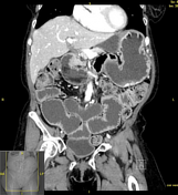
A constellation of findings related to scleroderma (gastrointestinal manifestations).
- dilated fluid-filled lower oesophagus.
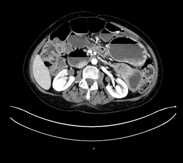
- dilated proximal to mid-small bowel segments, measuring up to 6.1 cm in calibre with a transition to non-dilated distal small bowel. Wall thickening of the jejunum with tightened jejunal folds - hidebound sign (bowel). No bowel fistula nor sinus tract.
Other findings:
- small right pleural effusion. Associated minimal smooth pleural thickening. The heart is normal in size.
- the liver, spleen, and adrenal glands are unremarkable. S/p cholecystectomy. No biliary duct dilatation.
- cystic lesions of the pancreatic head and uncinate process, measuring up to 1.8 cm. Increased enhancement of pancreatic uncinate process and posterior head without focal enhancing lesion. This may represent side-branch intraductal papillary mucinous neoplasms. No main pancreatic duct dilatation.
- 4 mm non-obstructing stone within the lower pole of the right kidney.
- the abdominal aorta is normal in calibre. Atherosclerotic vascular disease. Incidental retroaortic left renal vein.
- no enlarged abdominal lymph nodes. No abdominal free fluid or loculated fluid collections. Postoperative changes of the anterior abdominal wall.
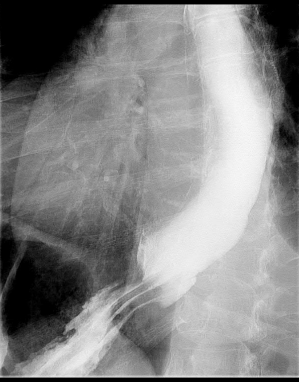
Oesophageal dilation and delay in oesophageal emptying due to stricture at the gastrooesophageal junction.
Case Discussion
The case exhibits characteristics of chronic systemic sclerosis on CT and esophagram, including hidebound sign (bowel), oesophageal dilatation, and lower oesophageal sphincter incompetence.
This case was submitted with supervision and input from:
Frank Chen, M.D. (Attending Radiologist) & Dillon Brown, M.D. (Resident Radiologist)
Mayo Clinic, Jacksonville
Department of Diagnostic and Interventional Radiology


 Unable to process the form. Check for errors and try again.