
Presentation
Routine breast imaging
Patient Data
Age: 50 years
Gender: Female

Download
Info

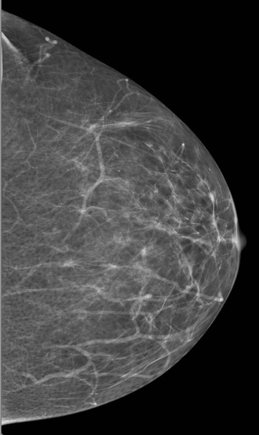
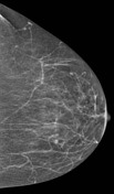
Small lesion has now appeared in the medial breast
From the case:
Screening mammogram - lobular carcinoma
Download
Info
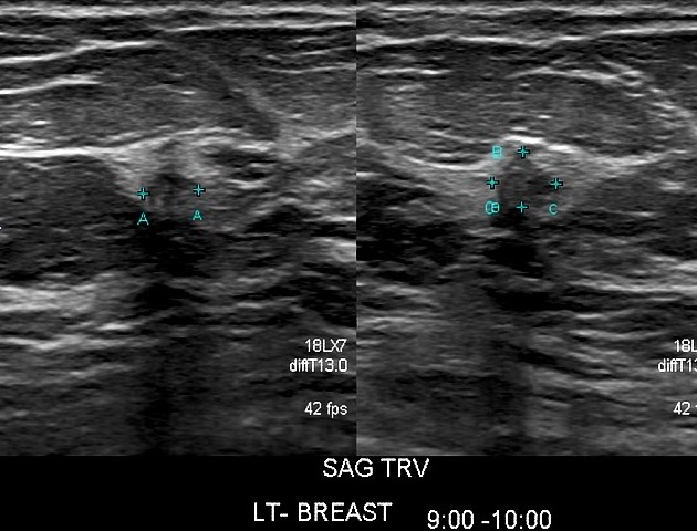
Small solid lesion of 4 mm confirmed on ultrasound.
Case Discussion
The value of using previous studies for comparison. It is quite exceptional to be able to biopsy a 4 mm lesion. Review what "overdiagnosis" is. Reading of screening studies should be done in isolated reading areas. If your reading environment is a generally busy, noisy area with distractions and phone calls, you will miss these lesions.
Confirmed lobular carcinoma.


 Unable to process the form. Check for errors and try again.