
Presentation
History previous mediastinal mass. PET-CT also showed pancreatic lesions. MRI for follow-up.
Patient Data


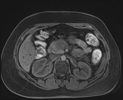
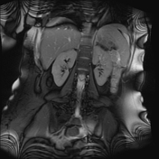
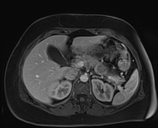

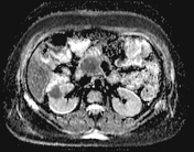
The MRI shows a partially imaged mass in the right lower anterior medisatium which shows significant diffusion restriction and post contrast enhancement. Enlarged lymph node in the right pericardial fat pad is noted.
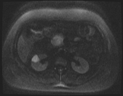
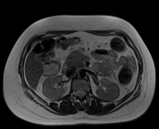
There also a number of hypoenhancing masses with the pancreas and both kidney which also show strong diffusion restriction.
No significant intraabdominal lymphadenopathy. SImple benign cyst noted in the spleen which is normal in size.
Case Discussion
The images features are suggestive of a pattern of secondary lymphoma supported by the strong diffusion restriction.
Renal involvement occurs in 3–8% of patients with lymphoma; the kidney is the most commonly involved part of the genitourinary tract 1.
Bone marrow aspirate and mediastinal mass biopsy in thise case showed Diffuse Large B-Cell Lymphoma (DLBCL).


 Unable to process the form. Check for errors and try again.