
Presentation
IV drug abuser developed fever, dyspnea and palpitation, cardiac echocardiography revealed vegetations on cardiac valves
Patient Data
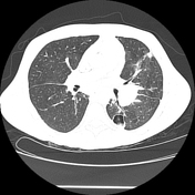


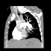
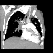

There is complete occlusion of the proximal right pulmonary artery with soft tissue density thrombus that extends to the ascending and descending branches. On the left side, the descending branch of left pulmonary artery shows two aneurysmal dilatation surrounded by soft tissue density ? inflammatory tissue versus leakage. Lung window images reveal multiple variable size lung nodules with typical feeding vessel sign as well as air filled cavitary lesions mostly small abscesses.
Case Discussion
Complete occlusion of the right pulmonary artery is suggestive of chronic pulmonary embolism rather than acute form. The left side pulmonary aneurysms are likely to be infective as the patient had no history of any sort of vasculitis or pulmonary hypertension. Multiple pulmonary parenchymal septic emboli and abscesses are a sequel of IV drug abuse.


 Unable to process the form. Check for errors and try again.