
Presentation
Pain around the first metatarsophalangeal joint for 3 weeks after a trauma.
Patient Data
Age: 35 years
Gender: Female
From the case:
Sesamoid fracture

Download
Info
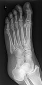
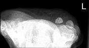
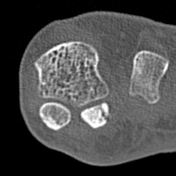
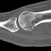
There is a vertical lucent line through the lateral sesamoid bone suggestive of a fracture.
From the case:
Sesamoid fracture

Download
Info

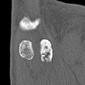
There is a comminuted and predominantly sagittal oriented fracture line through the proximal body of the lateral sesamoid bone. The fracture line is irregular and it appears to have sclerotic margins suggesting subacute fracture.
Case Discussion
Sesamoid fractures are not common and can be mistaken with bipartite sesamoid on plain x-ray.


 Unable to process the form. Check for errors and try again.