
Presentation
Longstanding shortening of the left leg. Fall. Fracture?
Patient Data
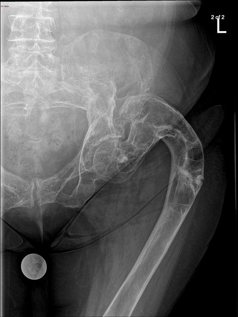
Shepherd crook deformity of the left hip as evidenced by a severe coxa varus angulation of the proximal femur. Underlying lucent expansile lesion in the femoral neck and proximal femoral diaphysis
Previous study from several...
Previous study from several yr ago
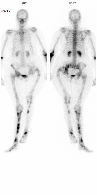
Multiple foci of increased tracers uptake in the long bones of the peripheral skeleton, left hip and pelvis. This is most pronounced and multifocal in the right tibia.
Shepherd crook deformity of the left hip.
Case Discussion
This is a case of polyostotic fibrous dysplasia. The most frequent sites of involvement are the femur, tibia and pelvis. This is giving rise to the classic shepherd crook deformity in the left hip.
Case submitted in conjunction with Dr Suman Bandhu.


 Unable to process the form. Check for errors and try again.