
Presentation
Known sickle cell disease patient was complaining of generalized abdominal pain.
Patient Data
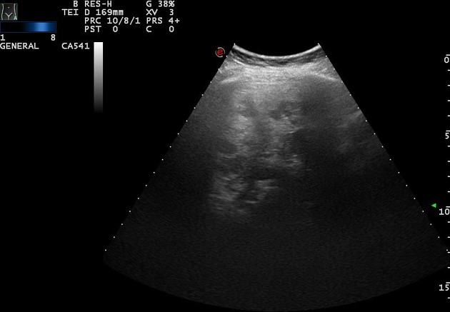
The spleen is not enlarged but has multiple variable sized hypo-echoic focal lesions.


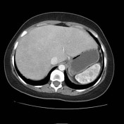
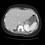
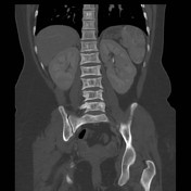
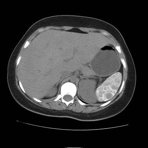
Liver is enlarged with no contoural irregularity. Two tiny hypodense focal lesions are noted at the right lobe (too small to characterize) likley simple liver cysts. Diffuse splenic hyperdensity and calcification is noted. Multiple hypodense focal lesions are scattered within splenic parenchyma showing mild enhancement at the post contrast series.Two tiny hyperdense left renal parenchymal foci are noted likely hemorrhagic cysts.
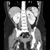
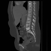
The visualized vertebral bodies showed decreased bone density with relatively decreased vertebral height giving H-shaped like appearance.
Case Discussion
Multiple splenic infarcts and bony changes in known chronic patient with sickle cell disease.


 Unable to process the form. Check for errors and try again.