Presentation
History of silica exposure, complaining of shortness of breath.
Patient Data
![](https://prod-images-static.radiopaedia.org/images/60548284/bc71b795573d540ecdbc97e410e7bc3a94f30a1694522c4cdc5a60c4a7c3dcad_thumb.jpeg)
![This study is a stack](/packs/stack-YQKLCKBI.gif)
![](https://prod-images-static.radiopaedia.org/images/60548815/7e92dabc533e71225d6ad7b0b11c898d2fc62ea6dda757b977b46feeaad74cce_thumb.jpeg)
![This study is a stack](/packs/stack-YQKLCKBI.gif)
![](https://prod-images-static.radiopaedia.org/images/60548969/80492aeaf1ec966fd2ced05601254a826bcb08f9652cf404bf8c8d2907460d47_thumb.jpeg)
![This study is a stack](/packs/stack-YQKLCKBI.gif)
![](https://prod-images-static.radiopaedia.org/images/60549020/bf81bb1e9dbe4a73c72640e9eb411f4e6bcae99258c7ffe26f091bcffad05cce_thumb.jpeg)
![This study is a stack](/packs/stack-YQKLCKBI.gif)
![](https://prod-images-static.radiopaedia.org/images/60549072/bbab78005e9c3e7e94b0ea68cfdee4d20f7bd7632a63ced7c40411fa31ceaf68_thumb.jpeg)
![This study is a stack](/packs/stack-YQKLCKBI.gif)
![](https://prod-images-static.radiopaedia.org/images/60549111/e91646590c5bfff7d8bcad69750b917336e82cc6e4f5a6f498fef26b87021423_thumb.jpeg)
![This study is a stack](/packs/stack-YQKLCKBI.gif)
![](https://prod-images-static.radiopaedia.org/images/60548284/bc71b795573d540ecdbc97e410e7bc3a94f30a1694522c4cdc5a60c4a7c3dcad_big_gallery.jpeg)
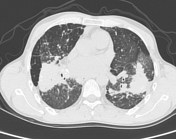
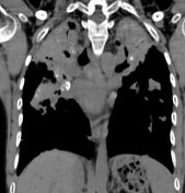
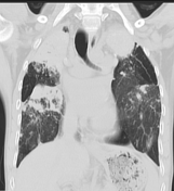
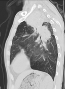
Extensive areas of bilateral coalescing perihilar and apical consolidation and fibrosis resulting in a mass-like appearance with architectural distortion and apical volume loss.
Numerous fine nodules throughout both lungs.
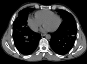
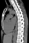
Numerous calcified mediastinal and hilar lymph nodes.
Mosaic attenuation and widespread interlobular septal thickening involving both lung fields.
Case Discussion
Consistent with patient's history of silica exposure, features are suggestive of complicated silicosis with progressive massive fibrosis.
To differentiate between progressive massive fibrosis and lung cancer we can use magnetic resonance imaging, where typically the lung cancer appears T2 hyperintense, whereas progressive massive fibrosis appears T2 hypointense (compared to skeletal muscle).