
Silicosis with progressive massive fibrosis
Citation, DOI, disclosures and case data
At the time the case was submitted for publication Frank Gaillard had no recorded disclosures.
View Frank Gaillard's current disclosuresPresentation
Progressive shortness of breath.
Patient Data


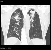
There is bilateral diffuse upper lobe reticular shadowing superimposed with occasional scattered mass like opacities.
1 case question available
Q: If the symptoms are progressive and there is no acute infection, what important occupational history needs to be elicited ? show answer
A: Occupations such as mining, quarrying, and tunneling

non-contrast

non-contrast

non-contrast

CT scan shows upper zone predominant mass-like scarring with calcification and volume loss. Hilar and mediastinal lymph node calcification also noted. No cavitary changes are seen. There is a left pleural effusion.
4 case questions available
Q: What is the most likely diagnosis ? show answer
A: Silicosis
Q: Is this simple or complicated silicosis ? show answer
A: Complicated silicosis
Q: Allowing for the absence of cavitatory and tree in bud changes, what would be an important differential diagnosis (as well as a potential complication) to consider? show answer
A: Pulmonary tuberculosis
Q: What incidental finding is present in the upper abdomen ? show answer
A: Pneumobilia
Case Discussion
Chest x-ray and CT scan of the chest demonstrate characteristic upper zone mass-like scarring with calcification and volume loss. Hilar and mediastinal lymph node calcification is also demonstrated. Features are in keeping with silicosis and progressive massive fibrosis (PMF).
References
- 1. Kim KI, Kim CW, Lee MK et-al. Imaging of occupational lung disease. Radiographics. 2001;21 (6): 1371-91. Radiographics (full text) - Pubmed citation
- 2. Chong S, Lee KS, Chung MJ et-al. Pneumoconiosis: comparison of imaging and pathologic findings. Radiographics. 2006;26 (1): 59-77. Radiographics (full text) - doi:10.1148/rg.261055070 - Pubmed citation
4 articles feature images from this case
409 playlists include this case
Public playlists
- chest x ray by Udithamala Priyanthi Ratnayake
- Nuha by Nuha homeida
- 2B VIVA MOCK 1 by Samuel T Gregson
- Upper lobe fibrosis by sananda haldar ◉
- Thorax by AT
- Viva 12 by Marios Zertalis
- Chest- General Approach by Annabelle Skelley
- Silicosis with progressive massive fibrosis by Özgen yılmaz
- Prova tórax by Cleverson Leitão
- chest viva by saba jaradat
- chest viva by BEN WONG
- SEP VIVA by BEN WONG
- FRCR 2b teaching 3 by Hassan Rehman
- Chest by Sourabh Malhotra
- Chest by Eugene Ng
- Chest e-film by Upuli Pahalawatta
- chest viva by Yu Xuan Kitzing
- Consolidation & Intersitial disease by Christopher L. Smoley
- BM - Chest by Blake Milton
- BPT Chest by Wayland Wang ◉ ◈
- Imanchest by Iman Fani
- MAK Chest by Michael Kreltszheim
- 02. interestitial by Gary Lee
- THX Occupational by Cary MacMillan
- P2 - Chest by Sachi Hapugoda
- My frcr 2b by Ahmed Samir
- Chest VIVAs VK1 by Kateryna Burlak
- RBWH hot seat pulmonary nodules by Annah Lane
- CXR by Kenneth Kwok
- Chest by Kayla Beck
- TGRS 4 by NKOSIYAZI HLABANO
- TRX neumoconiosis by Aram Ehsan Pernia
- Chest exam kist by Rajesh Pompapathy
- Chest with CT by Christian Acksteiner
- Revision - Chest by Aman Sandhu
- Multifocal Ill-defined opacities by Ryan McConnell
- MC Chest by Marianne Cossens
- JL Chest by Jacqueline Lubomski
- Chest by Paul Bui
- Plain film exam (CXR) by Andrew Panayiotou
- Chest 2 by Bob Ng
- Chest 2.0 by Sean Sweeney-Knapp
- Cxr by Pallavi Kamat
- Chest exam fodder by Gabrielle
- chest opacities by Bob Ng
- chest nov 2018 by Karina Dorfman
- Chest by Terrence Hui
- Plaman&Cardio-Vascular by Popovici Alexandra
- viva practice stars june 2024 3 by AHMED ELSHERBINI ELSABI
- Residents - Chest by Yaïr Glick ◉
- nezar chest by nezar shlaka
- Chest x ray by asim latif
- Chest X-ray patterns by ZUL KHAIRUL AZWADI BIN ISMAIL
- Registrar Random Viva 1 by Paul Bui
- Chest viva by Julia Williams
- Chest VIVA by Michael Le ◉
- Chest practice by Davina Bates
- Team FRCR Mock VIVA by Maha Elaassar
- Chest Viva by Manish99
- Core by Shekhar Khanpara
- chest by Manish Chug
- 2B VIVA 1 by Kishan Dissanayake
- Chest diffuse by Bob Ng
- Chest review by Ralph Nelson
- fibrosis by Madeleine Scicchitano ◉
- interstitial by Kutayba Adam
- Chest long cases by Udithamala Priyanthi Ratnayake
- eFilm 2 ZAK by Z Khan
- 2A Chest: 2 Patterns of lung disease by Daoud akhtar
- CHEST1 by Sean
- Chest by Gajan Surendra
- CHEST 1 BV by Bilal Vanlioglu
- Chest Xray Viva practice by Saneej Kanhirat
- eFilm reading 2018 (2) by Vicci du Plessis ◉
- phổi kẽ by Nguyen Thi Huyen
- chest by Manish Chug
- RAB Interstitial & Occupational by Arjuna Somanathan
- Chest-CVS 30-3-2022 by Mohamed R Nouh
- FRCR 2B Cases by Sherif Tharwat Gamal El Din ◉
- CHEST101 by Sherif Maccar ◉
- chest by R A A
- Chest by Whitney Graff
- Chest Review 1 by Timothy Jeffery
- Chest Tute 2 by Andrew Dixon ◉
- Chest board cases by Savannah Shortz
- Casinhos Tórax 15/12 by Flávio Murilo Ribeiro Bezerra
- Review by Christopher Lee
- Upper lobe fibrosis by Anastasia Tjan
- General 3B by Lisa Guion
- RS/Chest by Chinthaka Appuhamy
- YJL 2B Chest by YJ Lee
- CHEST 1 BV by Bilal Vanlioglu
- Chest 2 by Rod
- efilm2018(2) by Hassan Shoushtari Zadeh
- Cxr by Pallavi Kamat
- CHEST VIVA by Juliana Tsuruta ◉
- 2a by Prakruthi Venkatappa
- Core - chest by Csaba Ellák Siket
- Viva 1 by S Savaridas
- Tony Chen Chest exam 1 by Tony Chen
- Chest tut by Manish99
- 663 Consolidation & Interstitial by Eric Timothy Stefanowicz
- Thirsty Birds Pulmonary nodules by SM
- 2A Chest: 6 Diffuse lung disease by Daoud akhtar
- CHEST CASES by Mona Hiba
- Chest by shalabi
- GK - Chest - ILD by GLK
- Chest August 20 by Madeleine Scicchitano ◉
- Chest by Sameh Saied Ali ◉
- FRANZCR 02 Chest by AMIRSALEH JAFARI ◉
- Chest 2 April 22 ZAK by Z Khan
- Chest by Mai-Linh Le
- chest xray by SANDEEP KUMAR
- Thoracic FRCR 2B by Dr Feras Salhi
- chest by R A A
- cases 1 by Susantha indika Mahindawansha
- Chest by Joshua Taylor
- CxR Patterns by David Learmont-Walker
- eFilm Playlist 1 by Losing dePlot
- Chest Viva 2 by Saneej Kanhirat
- A 2021 long by Abdullah Hajar
- missed cases by Fahad Alabdulghani
- CHEST VIVA by Juliana Tsuruta ◉
- General -2 by Asmita Chugh
- Chest by Tamer Ghorab
- Chest by Mark Hall
- Chest test by Gary Lee
- UQ Radiology video tutorial: Chest: Interstitial lung disease by Craig Hacking ◉
- WDHB - Chest by Pieter Wood
- Pneumoconioses by Cleverson Leitão
- Buoi 23- Interstitial Pneumonia by Thuấn Nguyễn Hoàng
- Tute by Jayraj Singh Bhatti
- Chest - Clinical Conditions - Interstitial by Charlie Handley
- exam cases by Michael Ayeni
- Viva set 1 by Clement Chan
- Chest by Sean Lee ◉
- Chest by Nicholas Chen
- FRCR VIVA-Multisystem by Harshitha Shanbhag
- Chest 3 - Diffuse lung disease by Siobhan Lee
- Chest 3 by N Seth
- 170123 by Luke Hilliar
- Chest by Scott Mitchell
- Chest by Sean Lee ◉
- torax nodules by Aram Ehsan Pernia
- Dr Abid cases by Abid Hussain
- CHEST by wael
- 2B: Chest by Daoud akhtar
- FRCR 2 by Mohamed Salahaldin Elroos
- chest and cvs by Neelam Maharjan
- Chest by Jonathan Bong
- interstitial lung disease and toxic by Fatima Abdelmoniem Abdelrahman Ragab
- Chest 8-4-2023 by Mohamed R Nouh
- Chest 14-4-2023 by Mohamed R Nouh
- Random by Jithin Mohan
- FVW by Liana Franciscatto
- Interesting Cases by Raj Sohawon
- CXR by Ding Yi Zhang
- 2B CXR by GARETH MONTGOMERY
- Chest set pieces by Joshua Yap ◉ ◈
- Chest VIVA Set 1 by Cameron Grant
- CHEST by wael
- CHEST by wael
- CHEST by wael
- chest viva 1 pm by saba jaradat
- Thorax 12 - Apical masses by Praveen Samarawickrama
- Chest by Abdullah Hajar
- Haha by VAIBHAVKUMAR M VEKARIA
- Amila sir class play list by Udari Abeykoon
- Resp part 2 cases - Kevin Dunne by Kevin Dunne
- силикоз by Alla ivanova ivanova
- FRANZCR Chest by Sean Finnegan
- CHEST FRCR by Noha Aboelenin
- M chest by Maria Javed
- nezar chest by nezar shlaka
- RASHED 2B CHEST by Mahmoud Rashed
- CHEST CASES by Mohammed Naif AlJuraysi
- 2b by Mohamed Salahaldin Elroos
- chest by Tamer
- Chest cases by Malika Dhananjani Kumari
- Chest by Rasanayagam Mehatheepan
- VIVA 1 KP by Sarah Siaki Anane-Adusei
- CXR - lungs by Hari Prasath Sivakumar
- Chest by Dasun Kalugala
- Chest FRCR 2B by Rachel Iles Dillon
- Lung buddhi by Buddhi Niroshana Abeywickrama
- Chest by Elias Sachawars ◉
- Spotters 2/5 by Naveen Kumar
- Upper lung fibrosis by Hatem Al Jaafari
- Marwa Azab Approach chest by marwa
- Marwa Azab Approach chest by marwa
- Upper lobe fibrosis by reenagupta
- Diffuse reticulonodular shadowing by reenagupta
- Multiple calcified lung nodules by reenagupta
- Multiple calcified lung nodules by reenagupta
- Diseases chest by marwa
- ED/BPT chest x-rays by Mark Bishay ◈
- Resp Spotfires by Steven Clare
- Joh Curtis by marwa
- 2B Chest by James Coey
- Case review by mansur sha'bani fard
- CHEST /CVS 2 by Din Badshah
- Cxr by Hamza
- 15.0 CT Thorax Common pathology by Sahil Gulabkhan Malek
- thorax by Andra Vasc
- My cases by Nuwan Jayawardana
- chest day 3 by Quratulain Shah
Unlisted playlists
This case is used in 203 unlisted playlists.
Related Radiopaedia articles
Promoted articles (advertising)
How to use cases
You can use Radiopaedia cases in a variety of ways to help you learn and teach.
- Add cases to playlists
- Share cases with the diagnosis hidden
- Use images in presentations
- Use them in multiple choice question
Creating your own cases is easy.
ADVERTISEMENT: Supporters see fewer/no ads


 Unable to process the form. Check for errors and try again.