
Presentation
Acute on chronic shortness of breath.
Patient Data

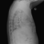
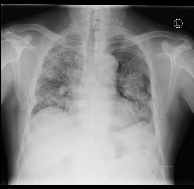
Lung architectural distortion with reticular opacities and mass-like lesions. Left-sided small pneumothorax. Enlarged paratracheal stipe due to lymphadenopathy.


There is a moderate left pneumothorax.
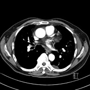
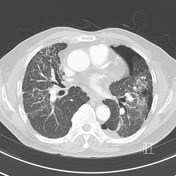
Severe upper zone predominant fibrosis with progressive massive fibrosis and centrilobular nodularity compatible with silicosis. Mildly enlarged mediastinal and bilateral hilar lymph nodes with scattered coarse calcification.
Mildly enlarged main pulmonary artery (3.1 cm) and dilation of right heart chambers, suggesting associated pulmonary arterial hypertension. Trace bilateral pleural fluid.
2 mm gallstone in the gallbladder neck. Imaged skeleton unremarkable.
Conclusion:
-Moderate left pneumothorax.
-Severe pulmonary infiltrates compatible with complicated chronic silicosis with progressive massive fibrosis.
-Stigmata of secondary pulmonary arterial hypertension
Case Discussion
This patient has a long-life work history as a stonemason. Features on imaging are those of progressive massive fibrosis secondary to silicosis and complicated by pneumothorax.


 Unable to process the form. Check for errors and try again.