
Presentation
Headache
Patient Data
Age: 30 years
Gender: Male
From the case:
Skull vault hemangioma

Download
Info
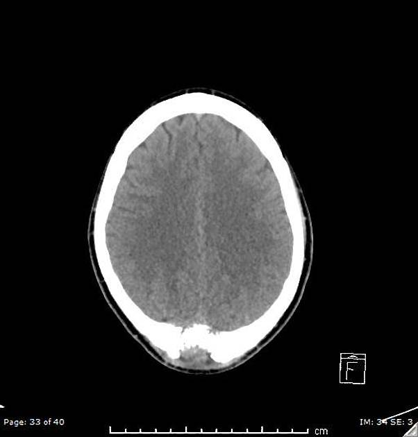
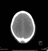
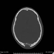
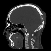
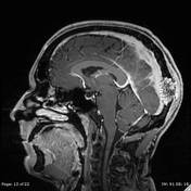
An expansile lesion in the occipital bone with a characteristic sunburst pattern of trabecular thickening. Erosions of both internal and external plates with associated internal and external tumor expansion.
From the case:
Skull vault hemangioma


Download
Info
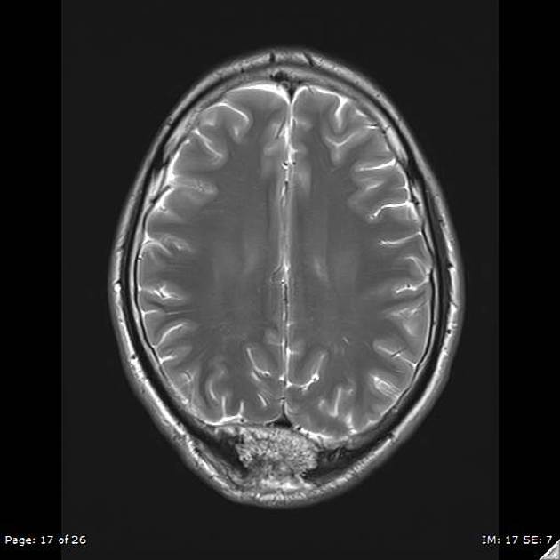
T2 shows hyperintense signal of the skull vault lesion.
T1 C+ shows enhancement of the lesion with epidural and subgaleal components.
Case Discussion
Skull vault hemangioma are benign, slow-growing, generally asymptomatic, vascular neoplasms. They represent 10% of benign neoplasms of the skull.


 Unable to process the form. Check for errors and try again.