
Presentation
Left frontal swelling for 7 years. Non-tender and slowly increasing in size.
Patient Data
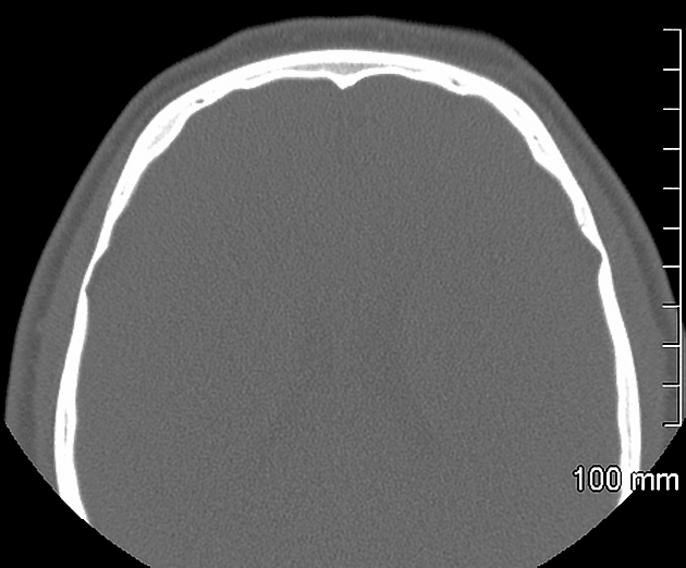
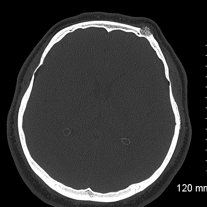
Small well-defined lesion with coarse trabeculations involving the outer table of left frontal bone measuring about 6 x 10 mm, associated with minimal swelling of the overlying soft tissues.
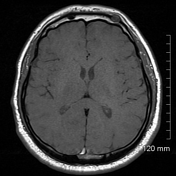
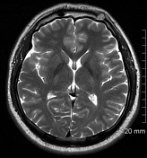
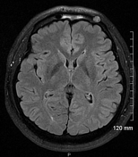
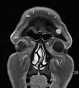
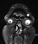
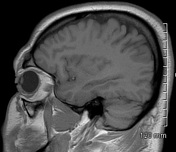
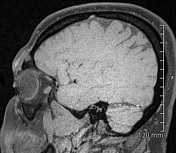
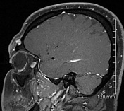
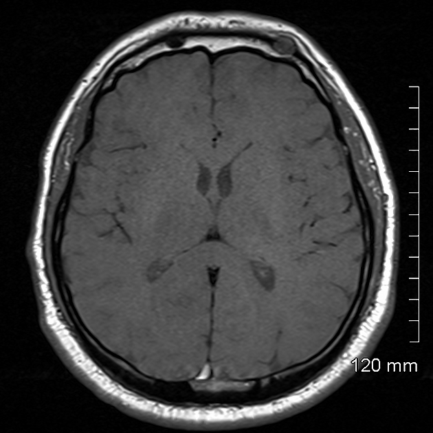
Small well-defined lesion involving the outer table of the left frontal bone. It is slightly hyperintense on T2 and isointense-hypointense on T1-weighted images relative to the bone marrow. It shows homogeneous enhancement on the post-contrast study.
The lesion has mildly increased in size and now it measures 10 x 15 mm, as compared to the previous measurement of 6 x 10 mm. It is associated with mild thickening and contour bulge in the overlying galeal soft tissues.
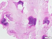
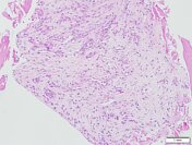
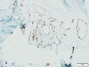
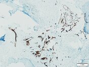

Capillary proliferation filling the bone trabeculae. CD34 highlights the vascular proliferation confirming the diagnosis of hemangioma.
Case Discussion
Slowly growing small well-defined lesion involving the outer table of the left frontal bone; the differential diagnosis for this lesion includes skull vault hemangioma, osteoma, eosinophilic granuloma and fibrous dysplasia.
The lesion was excised.
Procedure: Curettage of left frontal bone lesion.
Diagnosis: Skull vault hemangioma.


 Unable to process the form. Check for errors and try again.