
Presentation
Progressively worsening abdominal pain over past few weeks.
Patient Data
Age: 80 years
Gender: Male
From the case:
Small bowel perforation - ingested bone


Show annotations
Download
Info
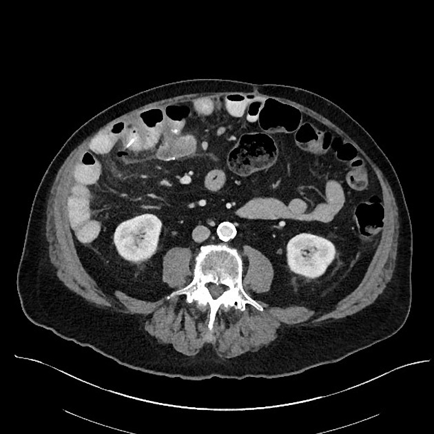
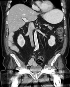
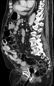
3 cm in length linear, dense foreign body within the loop of small bowel in the right mid abdomen, nearly traversing the wall. Adjacent to this are a few locules of extraluminal air with small amount of fluid.
Prior right hemicolectomy. No other acute findings.
Case Discussion
This case provides a great example of small bowel perforation due to foreign body. The appearance of the foreign body is characteristic of perforation by a thin bone fragment (most commonly a fish or chicken bone). Curiously, no foreign body was identified at the time of gross pathology, however, the CT findings of foreign body perforation are very convincing.


 Unable to process the form. Check for errors and try again.