
Presentation
Follow-up CXR post trauma 2 weeks ago
Patient Data
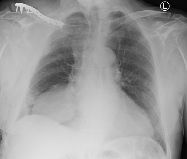
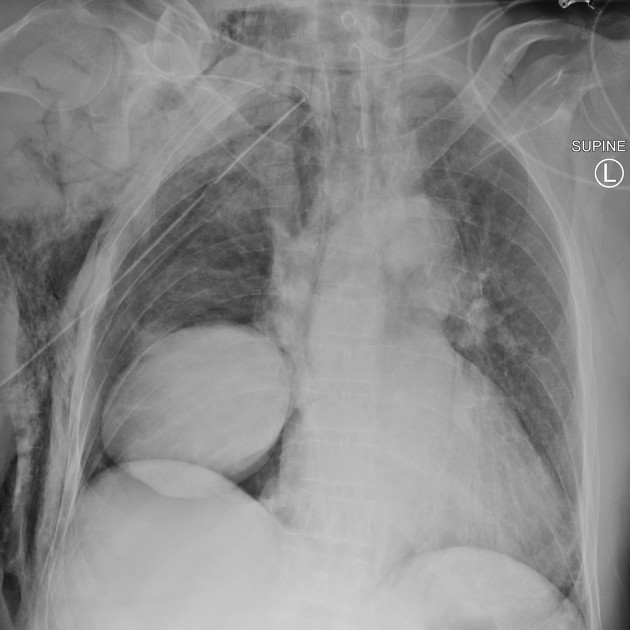
Well marginated rounded opacity in the medial right lower zone. No calcification or cavitation. The differentials for this lesion are broad based on the CXR findings and include: solitary fibrous tumor of the pleura, pericardial cyst, foregut duplication cyst, lung cancer, diaphragmatic hernia or less likely neurogenic tumor.
Soft tissue gas at the right base of neck and lateral chest wall. Right clavicle ORIF.
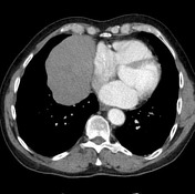

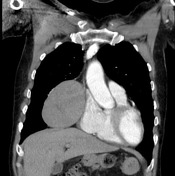
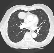
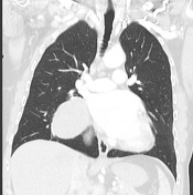

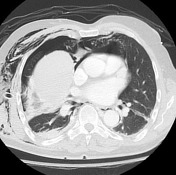
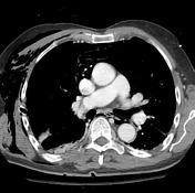
Well circumscribed 12cm solid pleural mass at the right cardiophrenic angle. There is mild indentation of the right atrium.
The lungs and pleural spaces are clear.
No mediastinal or hilar lymphadenopathy.
Taken at time of presentation post bicycle accident
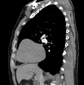
The right pleural mass is outlined by pneumothorax with very crisp margins.
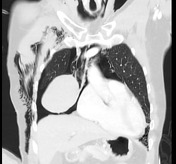

The right pleural lesion, including its pedicle, is outlined by the pneumothorax.
Large volume pneumomediastinum.
Right ICC in situ.
Case Discussion
Solitary fibrous tumor of the pleura is often attached by a pedicle (as seen in this case outlined nicely by the pneumothorax). They may be small (1 to 2 cm in diameter) or may completely fill the hemithorax.
Rarely, this tumor may be malignant, marked by pleomorphism, mitotic activity, necrosis, and large size (>10 cm). The tumor cells are positive for CD34 and STAT6 and negative for keratin which is the opposite of mesothelioma.


 Unable to process the form. Check for errors and try again.