
Presentation
Chest discomfort
Patient Data

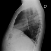
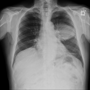
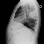
Frontal and lateral chest radiograph demonstrates a left paraspinal opacity that appears pleural based with an obtuse angle.
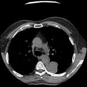


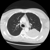
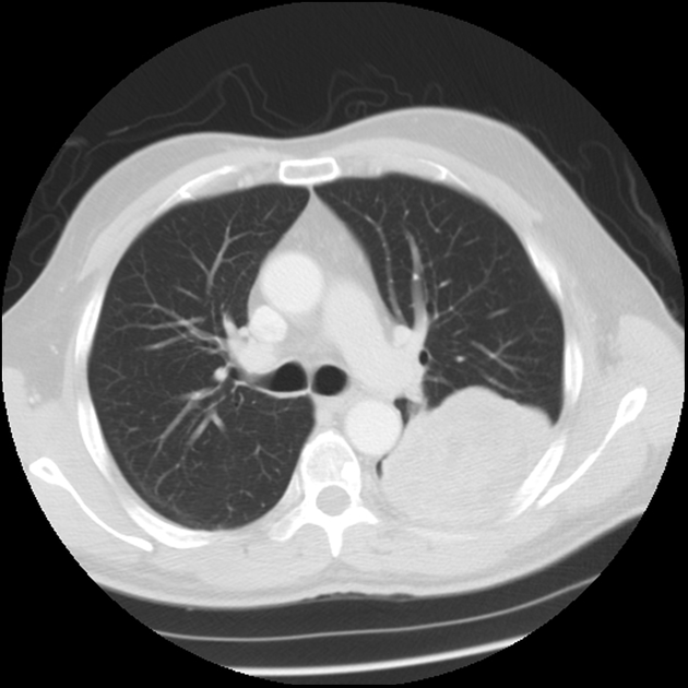
CT chest without contrast demonstrate a relatively well defined, lobulated pleural-based lesion at the left posterior lung.
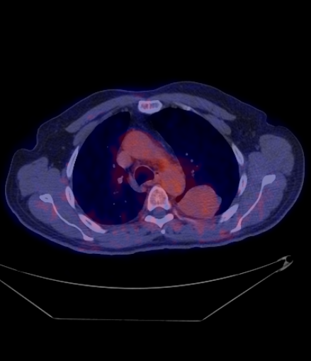
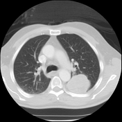
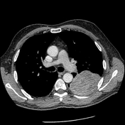
PET CT demonstrates that the lesion was not FDG-avid, with an SUV of 2.5
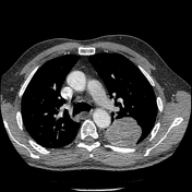

The lesion demonstrate interval increase in size as compared to the initial study. Heterogenous enhancement is evident.
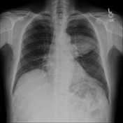
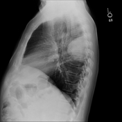

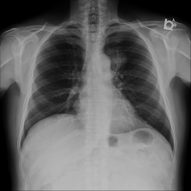
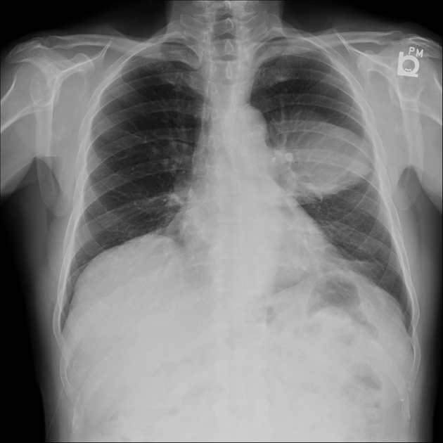
Frontal and lateral chest radiograph demonstrated marked increase in the size of a previously seen pleural-based lung lesion.

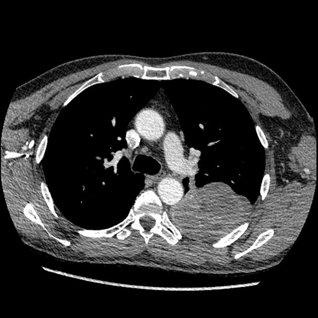
CT scan of the chest with contrast demonstrating interval increase in the size of the lesion with heterogenous enhancement. The lesion does not demonstrate any chest wall or vascular invasion.

Follow-up after 2 months demonstrate further increase in the size of the lesion.
Frontal and lateral chest radiograph after 1 month.
Case Discussion
Pathology: Pleural based spindle cell tumor with focal hypercellularity, moderate cytologic atypia, increased mitosis ( up to 10-12/10 HPF), and focal necrosis consistent with malignant solitary fibrous tumor of the pleura.
Solitary fibrous tumors are rare mesenchymal neoplasms that most commonly arise from the pleura, although they may also occur in the mediastinum and elsewhere in the body. They account for less than 5% of all pleural tumors and are not associated with asbestos exposure.
These tumors are slow growing. Patients can be asymptomatic or may present with local or systemic effects produced by the tumor.
Histologically, the majority of those tumors arise from the visceral pleura. Most of these tumors are low-grade neoplasms of variable cellularity and collagen content. Myxoid change and collagen degeneration may occur. Approximately 20% to 30% of solitary fibrous tumors are malignant, based on histologic criteria including high cellularity, high mitotic activity, and pleomorphism, as in our case.


 Unable to process the form. Check for errors and try again.