
Presentation
Right chest pain
Patient Data
Age: 55 years
Gender: Male
From the case:
Solitary fibrous tumor - pleura


Download
Info
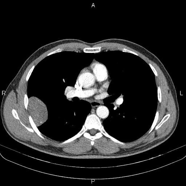
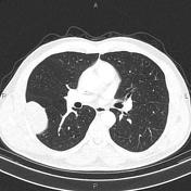
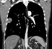
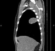
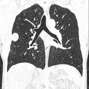
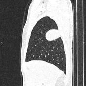
A 60×40 mm well-defined soft tissue density pleural- based mass with smooth margin and some internal calcifications is noted at mid portion of right hemithorax.
No chest wall invasion is noted. Additionally, a few subpleural nodules are seen at both lungs less than 8mm which seems to be fibrotic nodules. There are also several atelectatic bands scattered bilaterally.
Mild degenerative changes as osteophytosis are seen at the thoracic spine.
Case Discussion
Pathology proven pleural solitary fibrous tumor, also known as pleural fibroma, which is a rare benign pleural-based tumor that accounts for <5% of all tumors involving the pleura.


 Unable to process the form. Check for errors and try again.