
Presentation
Weakness of the lower limbs
Patient Data
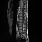

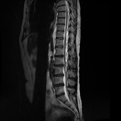
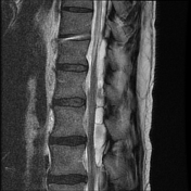
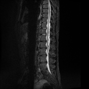
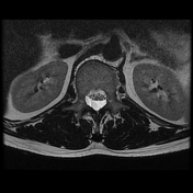

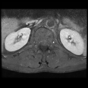

The MRI sequences demonstrate a septated posterior epidural cystic lesion of CSF signal on all sequences extending from T10-T11 to L2-L3 level with scalloping of the adjacent vertebral bodies and widening of the neural foramina. A mass effect is noted on the lower spinal cord and thecal sac which are displaced anteriorly. There is segmental atrophy of the spinal cord at T11 with area of high signal on T2/STIR indicating most likely a compressive myelopathy.
Case Discussion
MRI features of a spinal arachnoid cyst of posterior epidural location with compressive myelopathy of the lower spinal cord.
The spinal arachnoid cyst of epidural location is considered as a rare etiology of a compressive myelopathy 1. The most common reported site was at the lower thoracic region 1.
Additional contributor: A. Ramdani, MD


 Unable to process the form. Check for errors and try again.