
Presentation
Quadriplegia
Patient Data
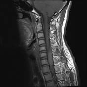

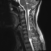
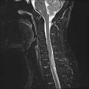
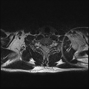
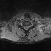
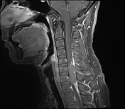
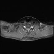
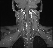

MRI of the cervical spine with contrast enhancement reveals a space-occupying lesion in the intraspinal - epidural on the right posterior side at the C6-C7 level. The lesion measures approximately 37 x 17 x 9 mm, with well-defined borders, causing significant compression and deformation of the cervical spinal cord at this level. It shows high signal intensity on T1W, STIR, and T2W sequences, with some scattered low-intensity areas on GRE. There is no contrast enhancement after injection.

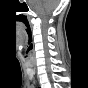
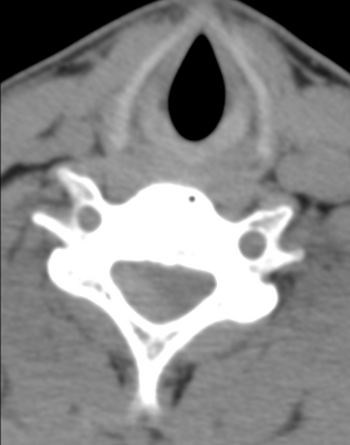
On non-contrast-enhanced CT, a lesion is observed in the cervical spinal canal at the C6-C7 level, with high density approximately 60 HU.
Surgical report:
the patient is intubated and positioned prone, with the Mayfield frame secured
an incision is made from the spinous process of C5 to the spinous process of D1
a partial laminectomy of C6 - C7 on the right side reveals an epidural hematoma extending from C6 to C7, coagulated and dark brown in color. The hematoma is completely evacuated. The bleeding source is from the epidural vascular system due to a vascular malformation. Hemostasis is achieved using bipolar cautery, supplemented with Spongel for additional bleeding control. After hematoma removal, the spinal cord is observed to be pulsating
a single epidural drain is placed
Case Discussion
Imaging findings are consistent with a spinal epidural hematoma. The patient subsequently underwent surgery, which confirmed the diagnosis, during which the hematoma was removed, relieving spinal cord compression. Postoperatively, clinical symptoms improved.


 Unable to process the form. Check for errors and try again.