
Presentation
Neck pain and upper limbs numbness and pain, more left-sided for 6 weeks.
Patient Data


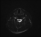
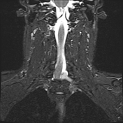
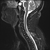
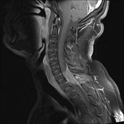
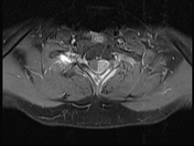
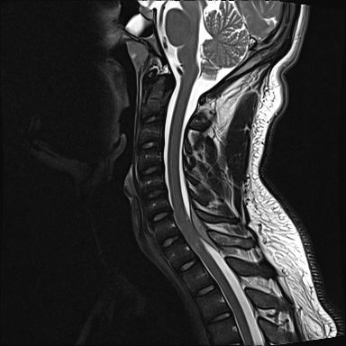
An ill-defined intraspinal extradural soft tissue lesion is seen at C7-T1 level eliciting isointense signal in T1 and hyperintense signal in T2 with homogeneous post contrast enhancement. The lesion extends into left C8 and T1 neural exit foramina. The spinous process and laminae of T1 vertebra show abnormal marrow signal intensity with heterogeneous post contrast enhancement.
Case Discussion
The differential diagnosis on imaging, in this case, is broad. Meningioma would typically have an enhancing dural tail and would not extend into the neural foramina. The neural foramina does not appear expanded which would have been in cases of schwannoma. Extramedullary hematopoiesis is unlikely as the abnormal bone marrow signal is limited to the level of the mass. Lymphoma and metastasis could also be considered in such case.
The main differentiating factor in this case is the abnormal marrow signal and bone involvement at the same level as the epicenter of the mass. Note that there is para-spinal soft tissue component along the laminae as well more on the left side.
Histopathology showed small blue round cell tumor consistent with Ewing sarcoma.


 Unable to process the form. Check for errors and try again.