


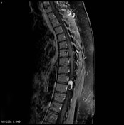
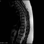
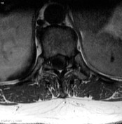
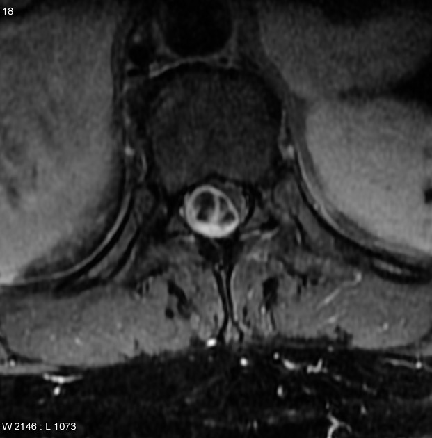
There is an extramedullary, intradural lesion, of lows T1 and high T2 signal, which is vividly, but heterogenously enhancing on post contrast images.
Note: This case has been tagged as "legacy" as it no longer meets image preparation and/or other case publication guidelines.
Case Discussion
Final Diagnosis:
Intradural T9-T10 tumor, resection: Schwannoma.
Gross Description:
The specimen [..] consists of a smooth-surfaced pink-yellowish nodule measuring 2 X 1.4 X 0.8 cm.
Microscopic Description:
[...] neoplasm with the typical histologic features of schwannoma. The tumor cells are arranged in compact, interlacing bundles in the Antoni type A areas and in a more loose fashion in the Antoni type B areas. There is focal formation of Verocay bodies. Scattered throughout are thick-wall hyalinized blood vessels. Within the center of the neoplasm there are variably dilated cystic spaces, some of which contain blood. The tumor is well circumscribed and surrounded by a compressed fibrous tissue pseudocapsule.


 Unable to process the form. Check for errors and try again.