
Presentation
Huge mass in the neck and upper back.
Patient Data
CASE OF THE MONTH: This case was selected as the Case of the Month for August 2024.


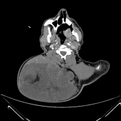
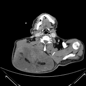
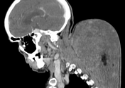
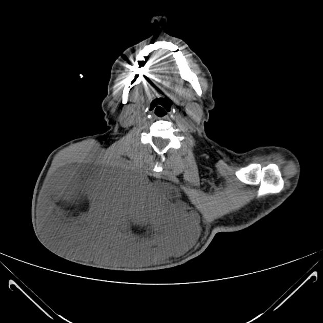
There is a huge lobulated subcutaneous mass in the posterior right neck and upper back. It extends downward from the level of C2 to D7, showing predominant heterogeneous enhancing soft tissue component with a few fat density areas, exerting mass effect on the underlying muscles, and abutting the superior edge of the right scapular process without bony destruction. The mass measures about 14.5 x 20 x 27 cm (AP, TS, CC).
The patient went on to have a resection.
Macroscopic pathology
The fixed specimen consists of a 2722 gm, 25 x 21 x 10 cm well-defined grey-brown, thinly encapsulated soft tissue mass. Sectioning reveals a solid, edematous myxoid cut surface with yellowish areas. Also submitted are 3 brownish fatty tissue pieces 12 x 7 x 0.8 cm.
Histology
The lesion is composed of a well-demarcated, thinly encapsulated proliferation of rather bland spindle cells intermixed with mature adipose tissue. It has extensive myxoid changes and many mast cells, as well as foci showing ropey collagen. No lip blasts or atypical hyperchromatic cells are seen.
Immunohistochemistry
Immunohistochemical stains including: MDM2, S-100 and CD34 reveals the spindle cells are strongly positive for CD34 with negative S-100 and MDM-2.
Final diagnosis
Case Discussion
Spindle cell lipoma is a benign lesion in which mature fat is replaced by collagen-forming spindle cells.
Spindle cell lipoma has a significant tendency to occur in the subcutaneous tissue of the posterior neck (as in this case), shoulder, and back.
Spindle cell lipomas are composed of a relatively equal ratio of fat and spindle cells, yet either component may predominate, and this variation in the ratio of fat and spindle cells is responsible for the wide spectrum of imaging features.
Differential diagnosis include liposarcoma or hibernoma.
Spindle cell lipoma is a benign lesion that is cured by local excision. It has never been reported to metastasize.


 Unable to process the form. Check for errors and try again.