
Presentation
Nonspecific upper abdominal pain. Lactate elevated on blood tests.
Patient Data
Age: 70 years
Gender: Male

Download
Info
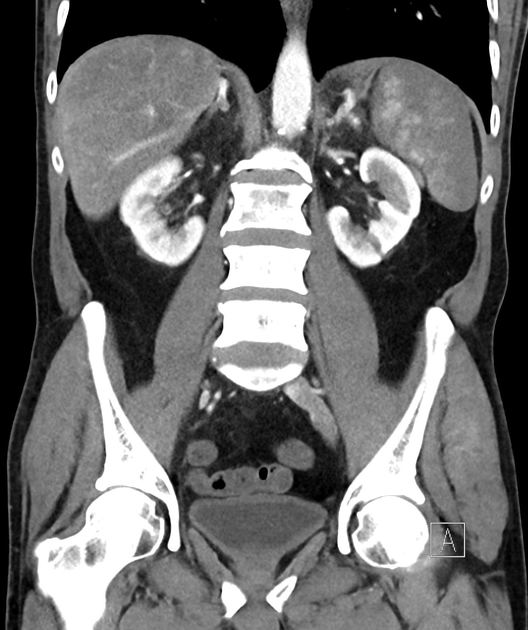
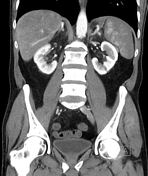
On these portal vein images, the spleen shows to be heterogeneously hypoperfused.
From the case:
Splenic infarct - liquefaction


Download
Info
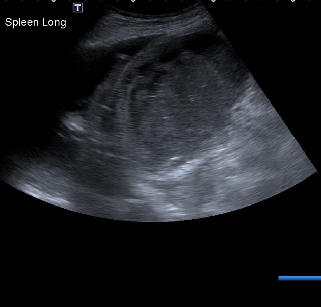
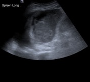
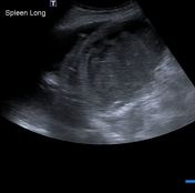
The spleen has heterogeneous echogenicity with an irregular contour in its upper posterolateral aspect with anechoic fluid in between these irregularities and the left diaphragm. A small pleural effusion is also noted on the left. Free fluid within the abdomen.
Case Discussion
This patient had CPK and LDH elevated. CT raised the suspicion for a perfusional disturbance involving the spleen, ultrasound 4 days later demonstrated heterogeneous echogenicity and irregular margins associated with subphrenic fluid. Features that are in keeping with splenic infarct with liquefaction.


 Unable to process the form. Check for errors and try again.