Presentation
Came of bicycle 24 hours ago - now pale, hypotensive, tender LUQ/flank pain.
Patient Data


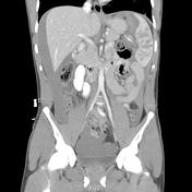
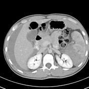
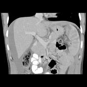
Post intravenous and oral contrast scans have been performed through the abdomen and pelvis.Severe splenic laceration, with no contrast enhancement demonstrated within the posteromedial half of the spleen.
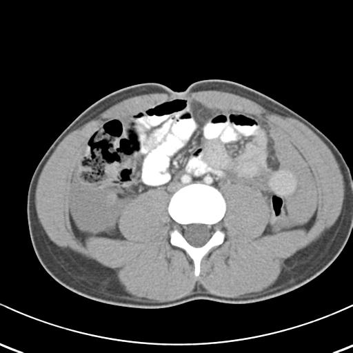
Within the anterolateral splenic fragment, a 1.2 x 0.7 cm hyperdensity is suggestive of a false aneurysm. Associated large amount of free intraperitoneal fluid / hematoma, tracking into the pelvis, paracolic gutters are around the liver.
Delayed phase demonstrates active bleeding into the nonenhancing splenic portion.No further intrabdominal visceral injury.
No fractures identified.
Case Discussion
Pseudoaneurysm formation is one of the complications of splenic laceration.


 Unable to process the form. Check for errors and try again.