
Presentation
Chest pain and shortness of breath since morning. No cough and sputum. No fever
Patient Data
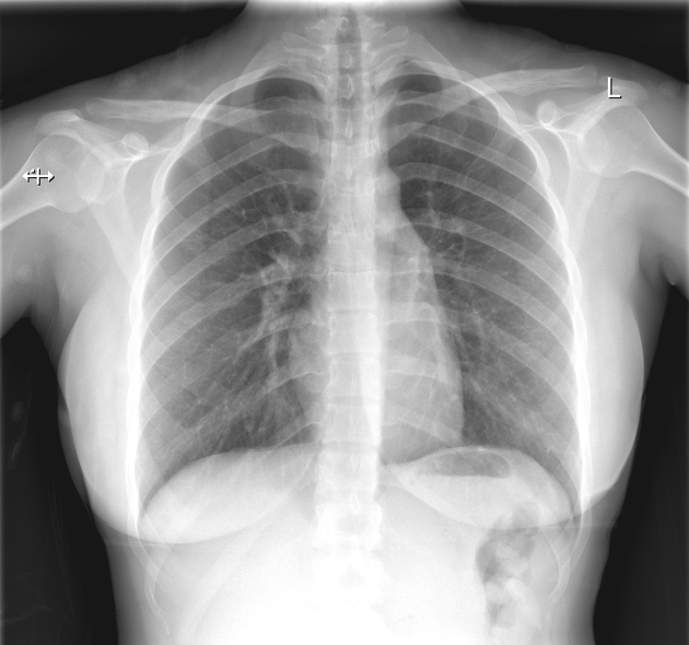
There are subtle linear paratracheal lucencies on both sides.
No visceral pleural line seen.
No pleural effusion.
No consolidation or collapse.
No osseous abnormalities.
Cardiac size appears within normal limits.
Lungs are clear


Air is outlining the trachea, oesophagus , left main bronchus and is extending to the neck around the carotid vessels.
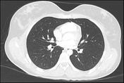
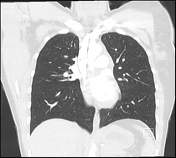
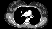
There are no pulmonary bullae, blebs or cysts.
No pneumothorax.
No pleural effusion.
Lungs are clear.
No mediastinal lymphadenopathy
No rib fractures.
Thoracic spine appears normal.
Case Discussion
This is a young patient presented with acute shortness of breath and chest pain without any history of trauma or cough.
The imaging revealed free air in the mediastinum.
Hence it is a case of spontaneous pneumomediastinum.
She did not gave any history of asthma, drugs, vomiting or Valsalva manoeuvres.
The symptoms were started after her morning exercise.
Subcutaneous emphysema was missing from the typical triad of thoracic pain, dyspnoea and subcutaneous emphysema.
Severe coughing or breath holding during strenuous exercise can lead to rupture of alveoli due to raised internal pressure.
An echocardiography will be technically difficult in such patients due to shadowing created by mediastinal free air.
She was admitted and treated with observation, bed rest ,oxygen and analgesics.
Monitored for complications such as infection or tension pneumothorax.


 Unable to process the form. Check for errors and try again.