
Presentation
Asymmetry in the cervical region.
Patient Data
Age: 1 year
Gender: Female
From the case:
Sprengel deformity


Show annotations
Download
Info
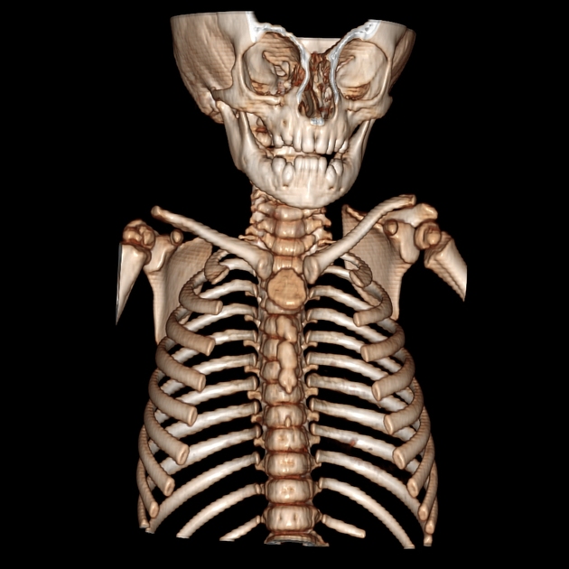
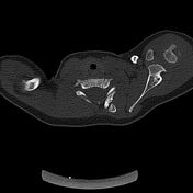
The left scapula is elevated with an accompanying omovertebral osseous bar arising from the fused laminae of C5-C6 to the middle part of the medial border of the scapula.
Midline posterior arch defect of C5 and C6 vertebrae consistent with type A posterior arch deformity.
The left vertebral lamina of C5 and C6 are fused.
Case Discussion
The observed features are indicative of Sprengel deformity, which is associated with Klippel-Feil syndrome and spina bifida.


 Unable to process the form. Check for errors and try again.