
Presentation
Yellowish vaginal discharge with recurrent episodes of bleeding.
Patient Data
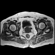

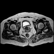
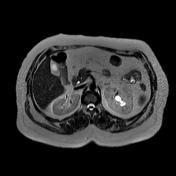
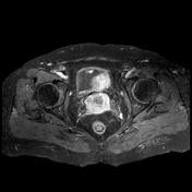
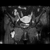

Irregular circumferential thickening of the cervical wall extends downward to involve the vagina with infiltration of the parametrium and few left side pelvic lymphadenopathies. There is also infiltration of the posterior wall of the urinary bladder and dilatation of the left ureter and hydronephrosis. The left kidney shows nephropathic changes.
Diagnosis: Cervical squamous cell carcinoma (pathology proven) T4N1M0 or FIGO IVa
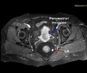
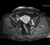
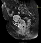
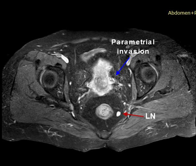
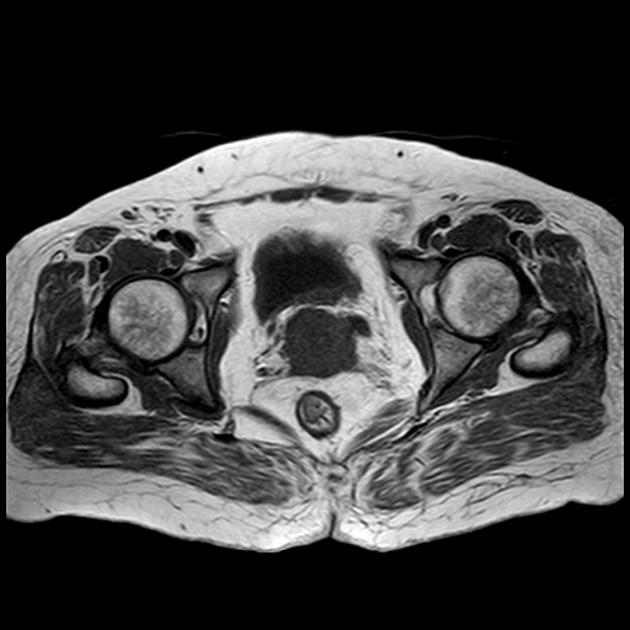
T1 postcontrast fat suppression images allow for better staging of cancer of the cervix.
Case Discussion
Cervical cancer staging is best assessed using MRI. With its superior tissue contrast, fat suppressed post contrast T1 weighted images can accurately demonstrate parametrial, vaginal and pelvic wall invasion as well as lymph node and ureteric involvement.


 Unable to process the form. Check for errors and try again.