
Presentation
Heavy smoker and alcohol abuse. Ulcerated lesion in the tongue.
Patient Data


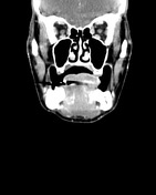
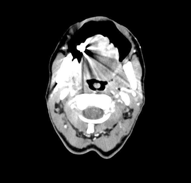
Enhancing ulcerated lesion within the anterolateral aspect of the tongue on the left. A few small left Ib lymph nodes are non-specific.
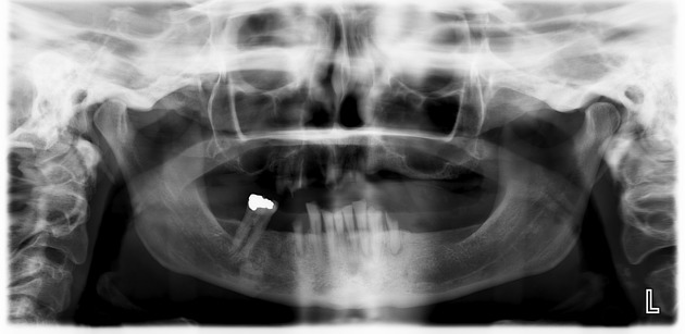
Poor dentition. No suspicious bone lesions.
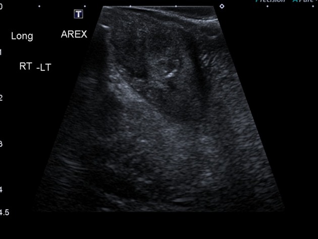
Heterogenous hypoechoic nodular lesion involving the anterolateral left aspect of the tongue, measuring 2.9 x 1.9 x 2.7 cm (across x deep x long).
Case Discussion
Punch biopsy of the tongue was performed:
MICROSCOPIC DESCRIPTION: Sections show stratified squamous mucosa and infiltrating squamous cell carcinoma with cords and nests of cells having keratin pearls and abundant intercellular bridges. There are numerous mitotic figures including atypical forms. The stroma is desmoplastic. There is no lymphovascular or perineural invasion identified. 2. Sections show a fragment of tissue predominantly composed of tumor of similar appearance to that seen in specimen 1 with a little benign, inflamed squamous epithelium at one end.
DIAGNOSIS: 1. Left anterior lateral tongue, biopsy: Squamous cell carcinoma. 2. Punch biopsy left anterior lateral tongue: Squamous cell carcinoma.


 Unable to process the form. Check for errors and try again.