
Presentation
Hematuria of 3 months duration. The patient is paraplegic with history of chronic bladder catheterization.
Patient Data
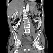

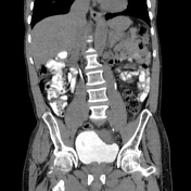
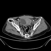
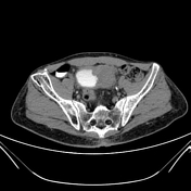

There is a heterogeneously enhancing mass with lobulated margins arising from the left superolateral wall of the urinary bladder extending through the wall with an exophytic component. There is invasion of the perivesical fat.
The pelvicalcyceal systems and ureters are within normal limits bilaterally.
There are multiple small bony fragments involving the right iliac bone likely post traumatic (past history of road traffic accident).
Diagnosis: Squamous cell carcinoma of the urinary bladder (histolopathology proven).
Case Discussion
Squamous cell carcinoma of the urinary bladder is uncommon but often seen in the setting of chronic bladder irritation. Risk factors include previous schistosomiasis infection, chronic bladder catheterization, bladder calculi and chronic urinary tract infections. The risk factor identified in this patient is chronic history of indwelling bladder catheter. Patient became paraplegic with history of urinary incontinence following a road traffic accident years ago.
HISTOPATHOLOGY
Microscopic examination of urothelial tissue shows sheets, nests and clusters of dyscohesive neoplastic squamous cells which are infiltrating the stroma. These cells are moderate in sizes and bear vesicular nuclei exhibiting marked pleomorphism, prominent nucleoli and mitotic figures of about 3/10hpf. No peripheral or lymphovascular invasion is seen. The base specimen contains these tumor cells and display effects of cauterization. Schistosoma ova are absent but marked infiltration by lymphocytes, plasma cells and neutrophils is seen.
DIAGNOSIS: INFILTRATING POORLY DIFFERENTIATED SQUAMOUS CELL CARCINOMA.


 Unable to process the form. Check for errors and try again.